Image processing apparatus and image processing method
a technology of image processing and image processing equipment, applied in the field of image processing apparatus and image processing method, can solve problems such as difficulty in acquiring an image at a high speed
- Summary
- Abstract
- Description
- Claims
- Application Information
AI Technical Summary
Benefits of technology
Problems solved by technology
Method used
Image
Examples
first exemplary embodiment
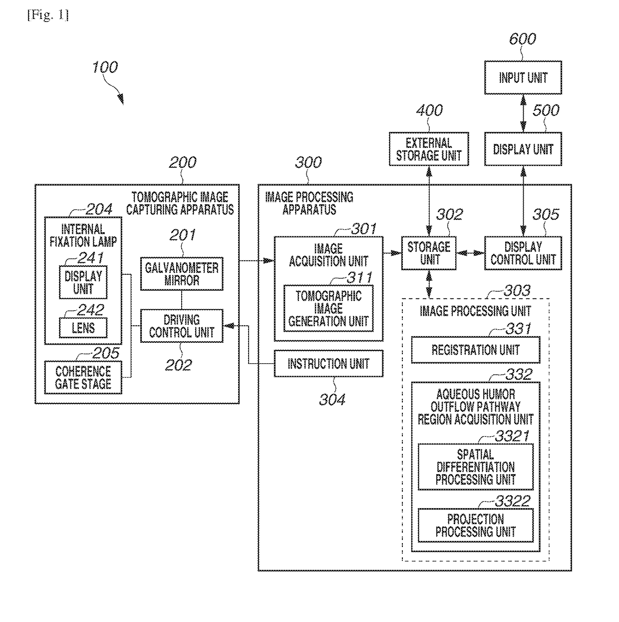
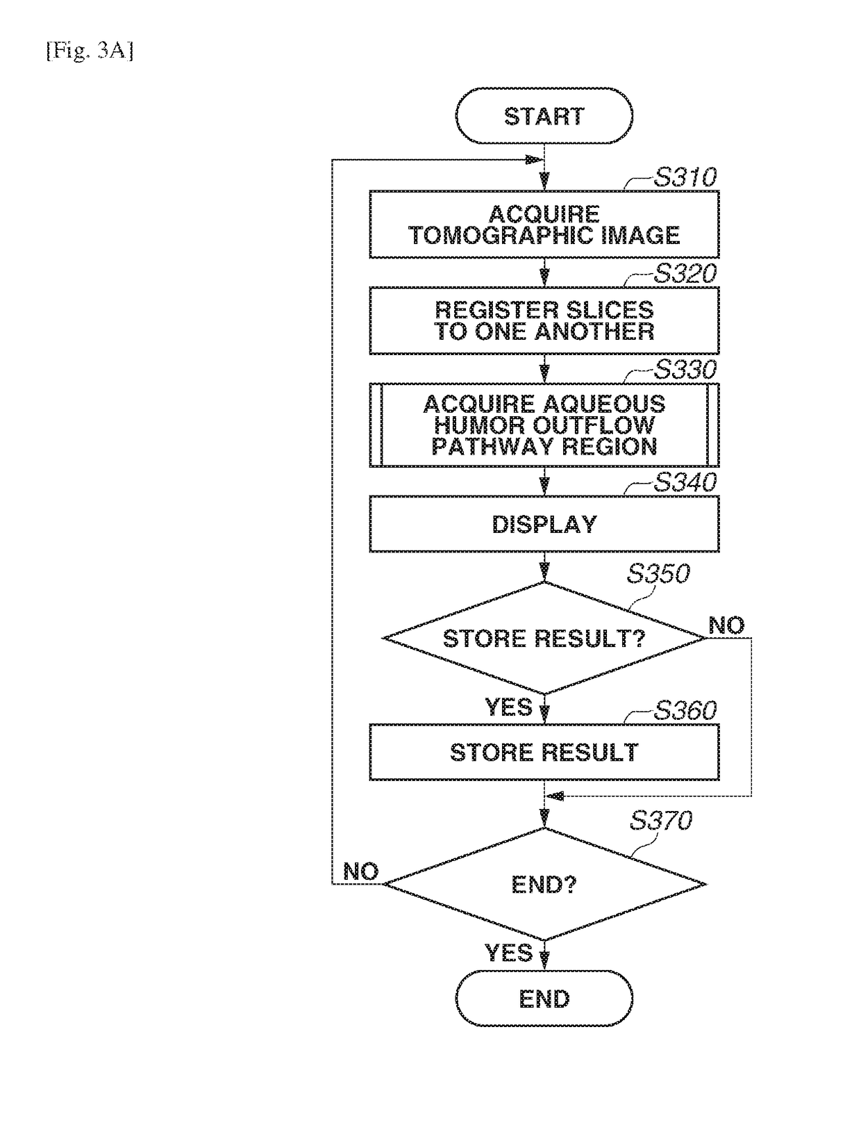
[0046]An image processing apparatus according to a first exemplary embodiment of the present invention will be described below. The image processing apparatus performs processing for differentiating a luminance value in a depth direction on the tomographic image of the anterior ocular segment that contains at least a deep scleral portion. Next, the image processing apparatus performs projection processing based on an amount of a variation in the luminance value in the depth direction with respect to different depth ranges of this differential image, thereby generating a group of projection images in which the aqueous humor outflow pathway region including the Schlemm's canal region SC and the collector channel region CC is emphasized in the different depth ranges. Further, the image processing apparatus binarizes each of these projection images based on a predetermined threshold value, thereby extracting a two-dimensional aqueous humor outflow pathway region.
[0047](Overall Configura...
second exemplary embodiment
[0089]An image processing apparatus according to a second exemplary embodiment of the present invention will be described below. The image processing apparatus generates an image in which a three-dimensional aqueous humor outflow pathway region including the Schlemm's canal region SC and the collector channel region CC is emphasized (drawn), by calculating a second-order differential of the luminance value in the depth direction with respect to the tomographic image of the anterior ocular segment that contains at least the deep scleral portion, and calculating an absolute value of this second-order differential value. Further, the image processing apparatus performs the extraction processing by binarizing this emphasized image.
[0090]The image processing system 100 including the image processing apparatus 300 according to the present exemplary embodiment is configured in a similar manner to the configuration according to the first exemplary embodiment, and therefore a description the...
third exemplary embodiment
[0106]An image processing apparatus according to a third exemplary embodiment of the present invention will be described below. The image processing apparatus identifies the Schlemm's canal region SC and the collector channel region CC from the aqueous humor outflow pathway region including the Schlemm's canal region SC and the collector channel region CC extracted with use of a similar image processing method to the second exemplary embodiment, and measures a diameter or a cross-sectional area of this aqueous humor outflow pathway region. Further, the image processing apparatus detects a lesion candidate region, such as stenosis, based on a statistical value of this measured value.
[0107]FIG. 7 illustrates a configuration of the image processing system 100 including the image processing apparatus 300 according to the present exemplary embodiment. The third exemplary embodiment is different from the second exemplary embodiment in terms of the image processing unit 303 including an id...
PUM
 Abstract
Description
Claims
Application Information
 - R&D
- Intellectual Property
- Life Sciences
- Materials
- Tech Scout
- Unparalleled Data Quality
- Higher Quality Content
- 60% Fewer Hallucinations
Browse by: Latest US Patents, China's latest patents, Technical Efficacy Thesaurus, Application Domain, Technology Topic, Popular Technical Reports.
© 2025 PatSnap. All rights reserved.Legal|Privacy policy|Modern Slavery Act Transparency Statement|Sitemap|About US| Contact US: help@patsnap.com


