Methods and systems for converting precursor cells into intestinal tissues through directed differentiation

a technology of directed differentiation and precursor cells, which is applied in the field of directed differentiation of precursor cells into intestinal tissues, can solve the problem that pluripotent stem cells alone cannot develop into and achieve the effect of reducing the number of fetal or adult animals

Active Publication Date: 2017-12-21
CHILDRENS HOSPITAL MEDICAL CENT CINCINNATI
View PDF0 Cites 9 Cited by
  • Summary
  • Abstract
  • Description
  • Claims
  • Application Information

AI Technical Summary

Benefits of technology

The patent describes a method for inducing the formation of intestinal tissue from precursor cells using specific signaling pathways. The method involves activating one or more of the Wnt, FGF, TGF-beta, shh, BMP, Notch, Hedgehog, LKB, and Par polarity signaling pathways in the precursor cell. The activated precursor cell is then cultured in vitro to form a 3-dimensional tissue structure. The intestinal tissue produced in this way can be used for various applications such as drug testing and tissue replacement therapy.

Problems solved by technology

Further, the pluripotent stem cells alone cannot develop into a fetal or adult animal because they lack the potential to contribute to extraembryonic tissue, such as the placenta.

Method used


Image

  • Methods and systems for converting precursor cells into intestinal tissues through directed differentiation
  • Methods and systems for converting precursor cells into intestinal tissues through directed differentiation
  • Methods and systems for converting precursor cells into intestinal tissues through directed differentiation

Examples


example 1

Directing Hindgut Development of PSCs

[0134]Maintenance of PSCs.

[0135]Human embryonic stem cells and induced pluripotent stem cells were maintained on Matrigel (BD Biosciences) in mTesR1 media. Cells were passaged approximately every 5 days, depending on colony density. To passage PSCs, they were washed with DMEM / F12 media (no serum)(Invitrogen) and incubated in DMEM / F12 with 1 mg / mL dispase (Invitrogen) until colony edges started to detach from the dish. The dish was then washed 3 times with DMEM / F12 media. After the final wash, DMEM / F12 was replaced with mTesR1. Colonies were scraped off of the dish with a cell scraper and gently triturated into small clumps and passaged onto fresh Matrigel-coated plates.

[0136]Differentiation of PSCs into Definitive Endoderm (DE).

[0137]Differentiation into Definitive Endoderm was Carried Out as Previously Described.

[0138]Briefly, a 3 day ActivinA (R&D systems) differentiation protocol was used. Cells were treated with ActivinA (100 ng / ml) for three...

example 2

Directing Hindgut Spheroids into Intestinal Tissue In Vitro

[0152]Directed Differentiation into Hindgut and Intestinal Organoids.

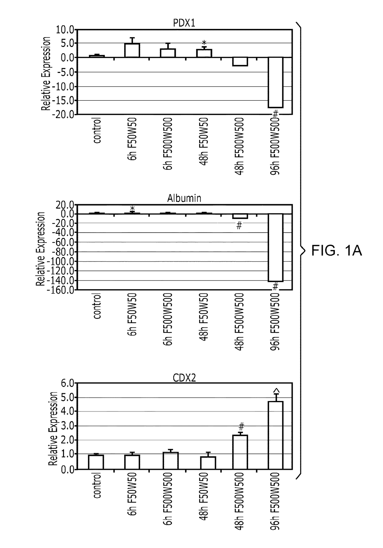
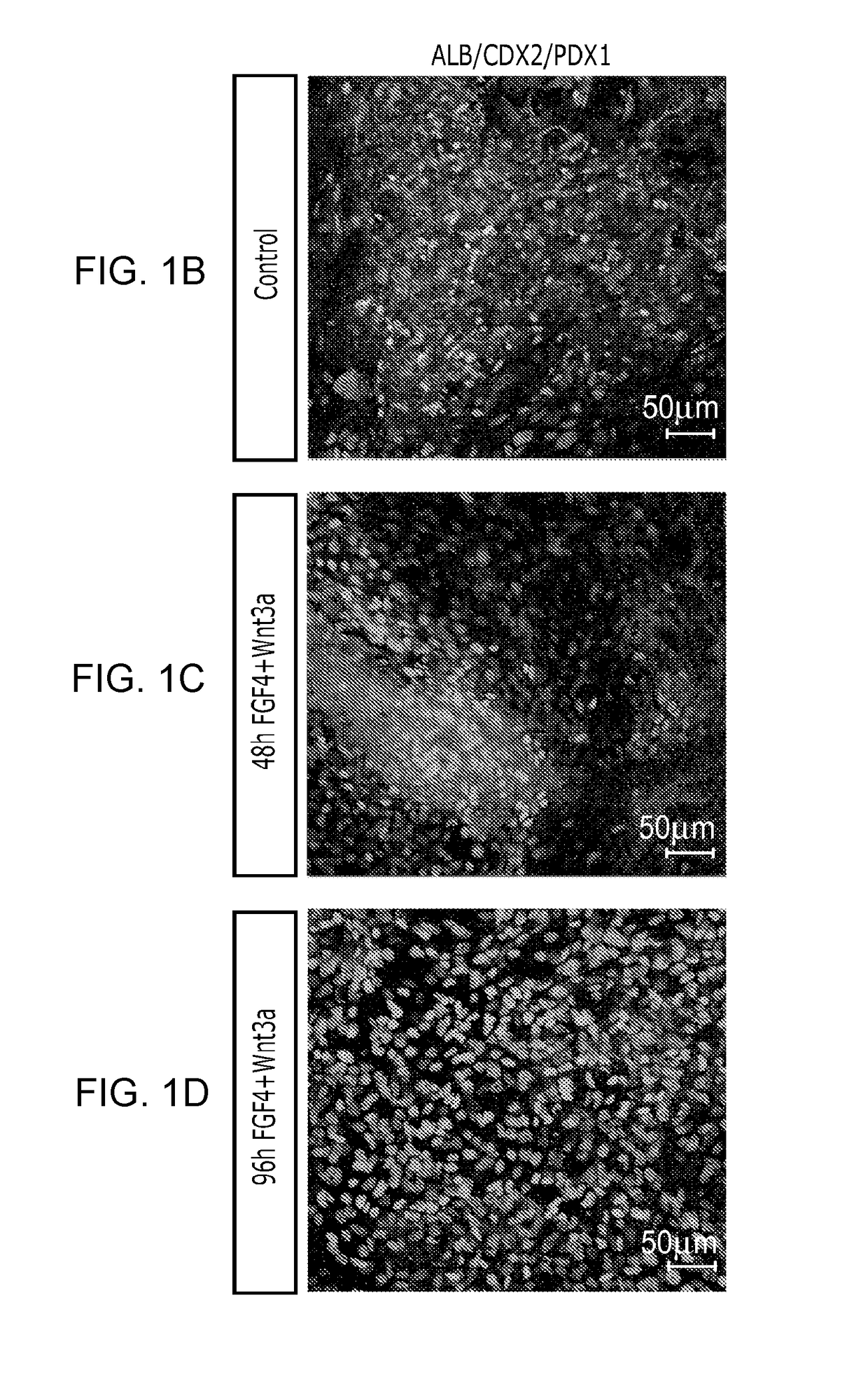
[0153]After differentiation into definitive endoderm, cells were incubated in 2% dFBS-DMEM / F12 with either 50 or 500 ng / ml FGF4 and / or 50 or 500 ng / ml Wnt3a (R&D Systems) for 2-4 days. After 2 days with treatment of growth factors, 3-dimensional floating spheroids were present in the culture. 3-dimensional spheroids were transferred into an in vitro system previously described to support intestinal growth and differentiation. Briefly, spheroids were embedded in Matrigel (BD Bioscience #356237) containing 500 ng / ml R-Spondin1 (R&D Systems), 100 ng / ml Noggin (R&D Systems) and 50 ng / ml EGF (R&D Systems). After the Matrigel solidified, media (Advanced DMEM / F12 (Invitrogen) supplemented with L-Glutamine, 10 μM Hepes, N2 supplement (R&D Systems), B27 supplement (Invitrogen), and Pen / Strep containing growth factors was overlaid and replaced every 4 days.

[0154]Dire...

example 3

Cytodifferentiation of PSCs into Mature Intestinal Cell Types

[0165]Between 18 and 28 days in vitro, it was observed that cytodifferentiation of the stratified epithelium into a columnar epithelium containing brush borders and all of the major cell lineages of the gut as determined by immunofluorescence and RT-qPCR (FIG. 4 and FIG. 11). By 28 days of culture Villin (FIG. 4a, a′) and DPPIV were localized to the apical surface of the polarized columnar epithelium and transmission electron microscopy revealed a brush border of apical microvilli indistinguishable from those found in mature intestine (FIG. 4d and FIG. 5). Cell counting revealed that the epithelium contained approximately 15% MUC2+ goblet cells (FIGS. 4a, a′), which secrete mucin into the lumen of the organoid (FIG. 11e), 18% lysozyme positive cells that are indicative of Paneth cells (FIG. 4b, b′) and about 1% chromogranin A-expressing enteroendocrine cells (FIG. 4c, c′; and FIG. 11). RT-qPCR confirmed presence of additio...


PUM


Abstract

The generation of complex organ tissues from human embryonic and pluripotent stem cells (PSCs) remains a major challenge for translational studies. It is shown that PSCs can be directed to differentiate into intestinal tissue in vitro by modulating the combinatorial activities of several signaling pathways in a step-wise fashion, effectively recapitulating in vivo fetal intestinal development. The resulting intestinal “organoids” were three-dimensional structures consisting of a polarized, columnar epithelium surrounded by mesenchyme that included a smooth muscle-like layer. The epithelium was patterned into crypt-like SOX9-positive proliferative zones and villus-like structures with all of the major functional cell types of the intestine. The culture system is used to demonstrate that expression of NEUROG3, a pro-endocrine transcription factor mutated in enteric anendocrinosis is sufficient to promote differentiation towards the enteroendocrine cell lineage. In conclusion, PSC-derived human intestinal tissue should allow for unprecedented studies of human intestinal development, homeostasis and disease.

Description

CROSS-REFERENCE TO RELATED APPLICATIONS[0001]This application is a continuation of U.S. patent application Ser. No. 13 / 695,887, filed Nov. 2, 2012, which is a U.S. National Stage Entry of PCT / US11 / 35518, filed May 6, 2011, which is based on and claims priority from U.S. Provisional Patent Application Ser. No. 61 / 332,178, filed May 6, 2010, the contents of which are incorporated by reference in their entirety for all purposes.STATEMENT REGARDING FEDERALLY-SPONSORED RESEARCH[0002]This invention was made with government support under GM072915, DK080823, DK084167, CA142826, DK083202, and HD007463 awarded by the National Institutes of Health. The government has certain rights in the invention.FIELD OF THE INVENTION[0003]The invention disclosed herein generally relates to methods and systems for converting stem cells into specific tissue(s) or organ(s) through directed differentiation. In particular, the invention disclosed herein relates to methods and systems for promoting definitive en...

Claims


Application Information

Patent Timeline
Patent Type & Authority Applications(United States)
IPC IPC(8): C12N5/071C12N5/077
CPCC12N2502/45C12N2502/02C12N5/0679C12N5/0661C12N2501/415C12N2501/155C12N2501/16C12N2501/385C12N2501/119
Inventor WELLS, JAMES M.ZORN, AARON M.SPENCE, JASON R.SHROYER, NOAH F.
Owner CHILDRENS HOSPITAL MEDICAL CENT CINCINNATI
Why Eureka
Try Eureka