Magnetic resonance thermometry during ablation

Active Publication Date: 2017-09-14
BIOSENSE WEBSTER (ISRAEL) LTD
View PDF7 Cites 3 Cited by
  • Summary
  • Abstract
  • Description
  • Claims
  • Application Information

AI Technical Summary

Benefits of technology

The invention relates to a method and apparatus for ablating tissue in a heart using a catheter with a position sensor and an electrode for ablation. The position sensor provides information about the location of the catheter in relation to the heart tissue being ablated, which helps to ensure that the ablation is being performed correctly. The method involves navigating the catheter to a target tissue, activating the ablation electrode, and acquiring magnetic resonance thermometry images at different positions to determine the temperature of the target tissue. The apparatus includes a flexible probe with a position sensor and a processor for controlling the magnetic resonance imager. The technical effects of the invention include improved accuracy and precision in ablating target tissue and reduced risk of complications.

Problems solved by technology

Cardiac arrhythmia, such as atrial fibrillation, occurs when regions of cardiac tissue abnormally conduct electric signals to adjacent tissue, thereby disrupting the normal cardiac cycle and causing asynchronous rhythm.
Procedures for treating arrhythmia include disrupting the areas causing the arrhythmia by ablation, as well as disrupting the conducting pathway for such signals.
A known difficulty in the use of radiofrequency energy for cardiac tissue ablation is controlling local heating of tissue.
There are tradeoffs between the desire to create a sufficiently large lesion to effectively ablate an abnormal tissue focus, or block an aberrant conduction pattern, and the undesirable effects of excessive local heating.
If the radiofrequency device creates too small a lesion, then the medical procedure could be less effective, or could require too much time.
On the other hand, if tissues are heated excessively then there could be local charring effects due to overheating.
Such overheated areas can develop high impedance, and may form a functional barrier to the passage of heat.
The use of slower heating provides better control of the ablation, but unduly prolongs the procedure.
However, for moving tissues, such as a beating heart, MRI images in sequence are generally not in registration with one another.
Accurate thermometry is not possible, because any observed phase change could be due, at least in part, to a movement in the target being imaged.

Method used


Image

  • Magnetic resonance thermometry during ablation
  • Magnetic resonance thermometry during ablation
  • Magnetic resonance thermometry during ablation

Examples


example

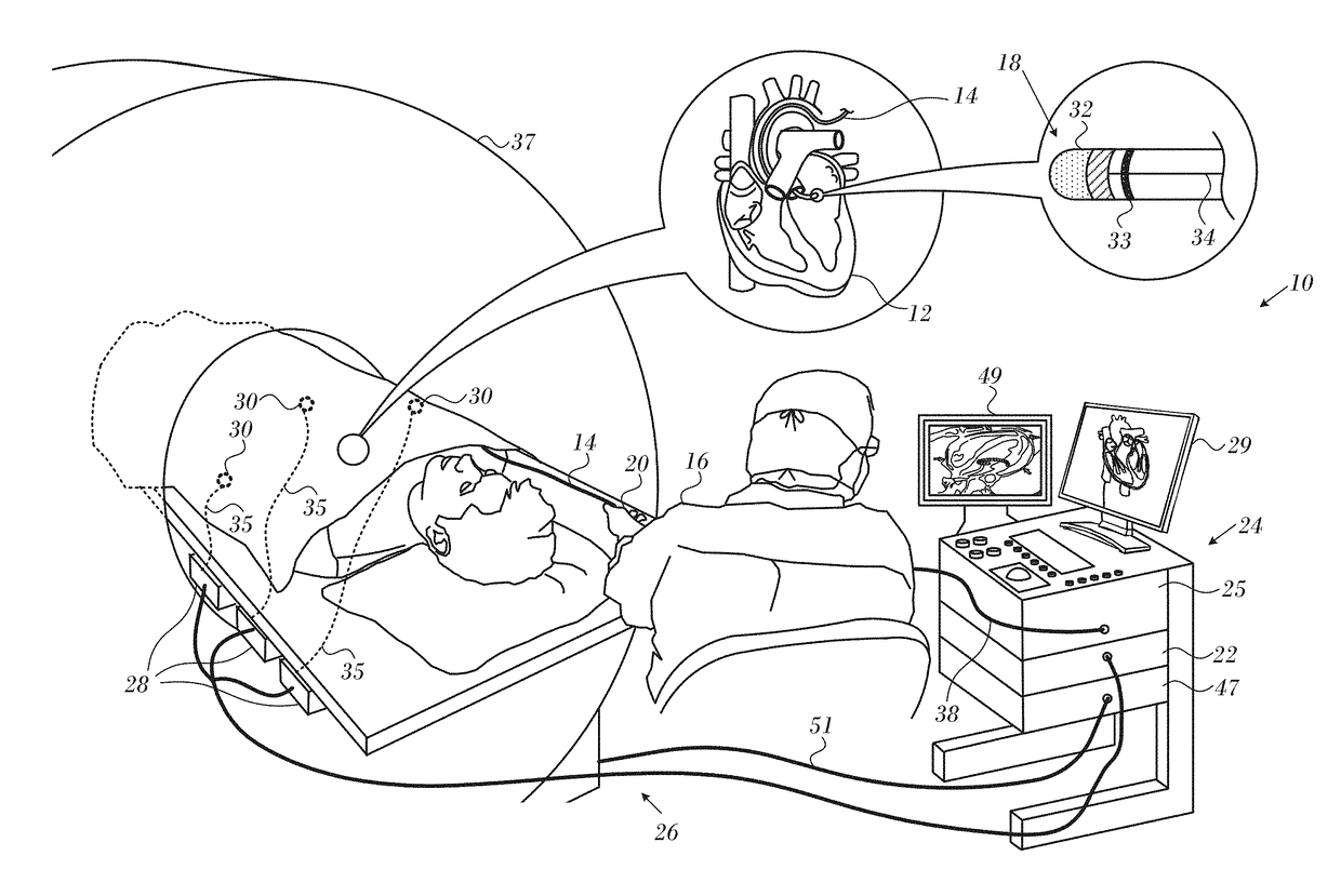
[0061]Reference is now made to FIG. 4, which is a composite diagram comprising two MRI thermography images 91, 93 showing typical results in a prospective cardiac ablation procedure in accordance with an embodiment of the invention. An increase in temperature 95 at ablation site 97 is noted in the later image 93, compared to temperature 99 in the earlier image 91. The operator can react to the measured temperatures 95, 99 by adjusting the power and / or duration of the ablation procedure as is known in the art.

[0062]Reference is now made to FIG. 5, which is a collection of two MRI images 101, 103 that are suitable for MRI thermography according to an embodiment of the invention. The images 101, 103 were obtained from data acquired in one slice in accordance with an embodiment of the invention. Images 101, 103 are an amplitude and a phase image, respectively. Ablation site 105 is indicated on both images.


PUM


Abstract

Thermography of an ablation site is carried out by navigating a probe into contact with target tissue in the heart, obtaining a first position of a position sensor in the probe and acquiring a first magnetic resonance thermometry image of the target tissue. The method is further carried out during ablation by iteratively reading the position sensor to obtain second positions, and acquiring a new magnetic resonance thermometry image of the target tissue when the distance between the first position and one of the second positions is less than a predetermined distance. The images are analyzed to determine the temperature of the target tissue.

Description

CROSS-REFERENCE TO RELATED APPLICATIONS[0001]This Application claims the benefit of U.S. Provisional Application No. 62 / 305,026, which is herein incorporated by reference.COPYRIGHT NOTICE[0002]A portion of the disclosure of this patent document contains material that is subject to copyright protection. The copyright owner has no objection to the facsimile reproduction by anyone of the patent document or the patent disclosure, as it appears in the Patent and Trademark Office patent file or records, but otherwise reserves all copyright rights whatsoever.BACKGROUND OF THE INVENTION[0003]1. Field of the Invention[0004]This invention relates to devices and methods for transferring non-mechanical forms of energy to or from the body. More particularly, this invention relates to magnetic resonance thermometry during cardiac ablation therapy.[0005]2. Description of the Related Art[0006]The meanings of certain acronyms and abbreviations used herein are given in Table 1.TABLE 1Acronyms and Abb...

Claims


Application Information

Patent Timeline
IPC IPC(8): A61B34/20G01R33/48A61B18/14G01R33/28A61B5/055A61B5/06
CPCA61B34/20A61B5/055A61B5/065A61B18/1492G01R33/287A61B2034/2051A61B2018/00351A61B2018/00577A61B2034/2065A61B2090/3954G01R33/4804A61B18/12A61B18/14A61B2018/00595A61B2018/00791A61B2018/1467G01R33/50G01R33/5605G01R33/56341A61B2090/374
Inventor GOVARI, ASSAFALTMANN, ANDRES CLAUDIOEPHRATH, YARONGLINER, VADIMDROR, EYAL
Owner BIOSENSE WEBSTER (ISRAEL) LTD
Why Eureka
Try Eureka