
Endoscope system and light source device

a technology of endoscope and light source, which is applied in the field of endoscope system, can solve the problems of low color separability, deterioration of reproducibility of color, and achieve the effect of improving the color separability and the visibility of the superficial blood vessels

Active Publication Date: 2017-05-11
FUJIFILM CORP
View PDF5 Cites 3 Cited by
  • Summary
  • Abstract
  • Description
  • Claims
  • Application Information

AI Technical Summary

Benefits of technology

This approach enhances the color separability and visibility of superficial blood vessels by optimizing the light ratio and signal processing, leading to improved contrast and clarity in narrowband light observation.

Problems solved by technology

The primary color type imaging device is used in an endoscope system that places importance on color, because of being superior in color reproducibility, though inferior in sensitivity, to the complementary color type imaging device.
On the other hand, the complementary color type imaging device, which is superior in sensitivity and inferior in color reproducibility to the primary color type imaging device, is used in an endoscope system that places importance on sensitivity.
This causes low color separability, and a blur of the superficial blood vessels due to the influence of scattered light deteriorates the visibility of the superficial blood vessels.
Therefore, the matrix operation cannot radically improve the color separability and the visibility of the superficial blood vessels.

Method used


Image

  • Endoscope system and light source device
  • Endoscope system and light source device
  • Endoscope system and light source device

Examples


Embodiment Construction

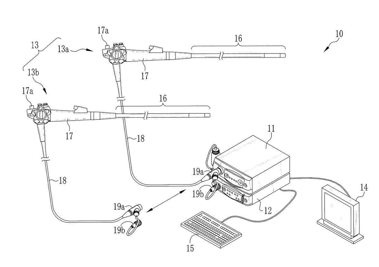
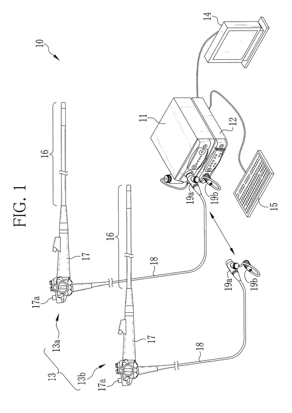
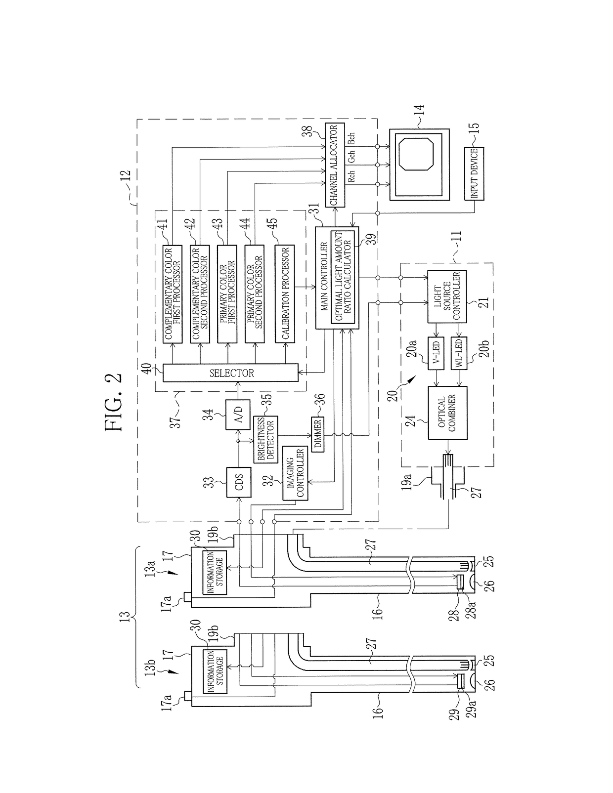
[0054]In FIG. 1, an endoscope system 10 is constituted of a light source device 11, a processor device 12, and electronic endoscopes 13 (hereinafter simply called endoscopes) detachably connected to the light source device 11 and the processor device 12. The light source device 11 produces illumination light and supplies the endoscope 13 with the illumination light. A distal end of the endoscope 13 is inserted into a human body cavity or the like to image the inside of the body cavity. The processor device 12 controls the imaging operation of the endoscope 13, and applies signal processing to an imaging signal obtained by the endoscope 13.

[0055]To the processor device 12, an image display device 14 and an input device 15 are connected. The image display device 14, being a liquid crystal display or the like, displays an image of an observation object inside the body cavity produced by the processor device 12. The input device 15, including a keyboard and a mouse, is used for inputtin...


PUM


Abstract

Violet narrowband light Vn and green narrowband light Gn produced by a light source device are supplied to a complementary color type endoscope, and simultaneously applied to an observation object. In a complementary color type imaging device, first mixed pixels and second mixed pixels, which sense both of the violet narrowband light Vn and the green narrowband light Gn, are read out. The light amount ratio Z of the violet narrowband light Vn to the green narrowband light Gn is set in such a range as to make the light amount of the violet narrowband light Vn larger than the light amount of the green narrowband light Gn, and make a signal value of the second mixed pixel higher than a signal value of the first mixed pixel.

Description

CROSS-REFERENCE TO RELATED APPLICATIONS[0001]This application is a Divisional of copending application Ser. No. 14 / 338,988, filed on Jul. 23, 2014, which claims priority under 35 U.S.C §119 to Japanese Patent Application No. 2013-202553 filed on Sep. 27, 2013. The above applications are hereby expressly incorporated by reference, in their entireties, into the present application.BACKGROUND OF THE INVENTION[0002]1. Field of the Invention[0003]The present invention relates to an endoscope system for performing narrowband light observation using a complementary color type imaging device, and a light source device used in the endoscope system.[0004]2. Description Related to the Prior Art[0005]In a recent medical field, diagnosis and treatment using an endoscope system, having a light source device, an electronic endoscope, and a processor device, are widely performed. The light source device produces illumination light and applies the illumination light to the inside of a human body cav...

Claims


Application Information

Patent Timeline
Patent Type & Authority Applications(United States)
IPC IPC(8): A61B1/06H04N5/225H04N9/04H04N9/07A61B1/00H04N5/378H04N9/12H04N9/77H05B33/08A61B1/04G02B23/24H04N5/372H04N23/12H05B44/00
CPCA61B1/0638H04N2005/2255G02B23/2469H04N5/2256H04N9/045H04N9/07H04N5/372H04N5/378H04N9/12H04N9/77H05B33/0845H05B33/0815A61B1/04A61B1/0684A61B1/00045G02B23/2484A61B1/00059A61B1/043H05B47/00A61B1/000094A61B1/044H04N23/555H04N23/56H04N25/136H04N23/843H04N25/134H04N23/12H04N25/71H04N25/75
Inventor KURAMOTO, MASAYUKI
Owner FUJIFILM CORP
Learn More