
Composition for imaging atherosclerosis and method for diagnosing atherosclerosis by using same

a technology of composition and atherosclerosis, applied in the field of composition for imaging atherosclerosis and a method for diagnosing atherosclerosis, can solve the problem of low manufacturing cost, achieve the effect of high diagnosis accuracy, effective diagnosis, and enable the diagnosis of atherosclerosis

Inactive Publication Date: 2017-03-09
KOREA UNIV RES & BUSINESS FOUND +1
View PDF0 Cites 0 Cited by
  • Summary
  • Abstract
  • Description
  • Claims
  • Application Information

AI Technical Summary

Benefits of technology

The present disclosure describes a composition that can accurately diagnose atherosclerosis, even for individuals with metabolic problems such as diabetes. Additionally, this composition is cost-effective and can be used for brain and heart atherosclerosis diagnosis. Overall, this invention offers an effective tool for identifying atherosclerosis and its associated risks.

Problems solved by technology

In addition, manufacturing cost is low compared with the existing imaging composition for diagnosis of atherosclerosis.

Method used


Image

  • Composition for imaging atherosclerosis and method for diagnosing atherosclerosis by using same
  • Composition for imaging atherosclerosis and method for diagnosing atherosclerosis by using same
  • Composition for imaging atherosclerosis and method for diagnosing atherosclerosis by using same

Examples


example 2

Preparation of Kit for Imaging Mannose Receptor

[0033]After adding 1 mL of the benzyl NOTA- and phenyl mannose-bound human serum albumin (13.6 mg / mL) to 0.3 mL of a sodium acetate buffer (0.5 M, pH 5.5), and transferring to each vial an amount corresponding to 1 mg of protein, the mixture was freeze-dried and stored at −70° C.

example 3

Preparation of 68Ga-Labeled Compound Using Kit for Imaging Mannose Receptor

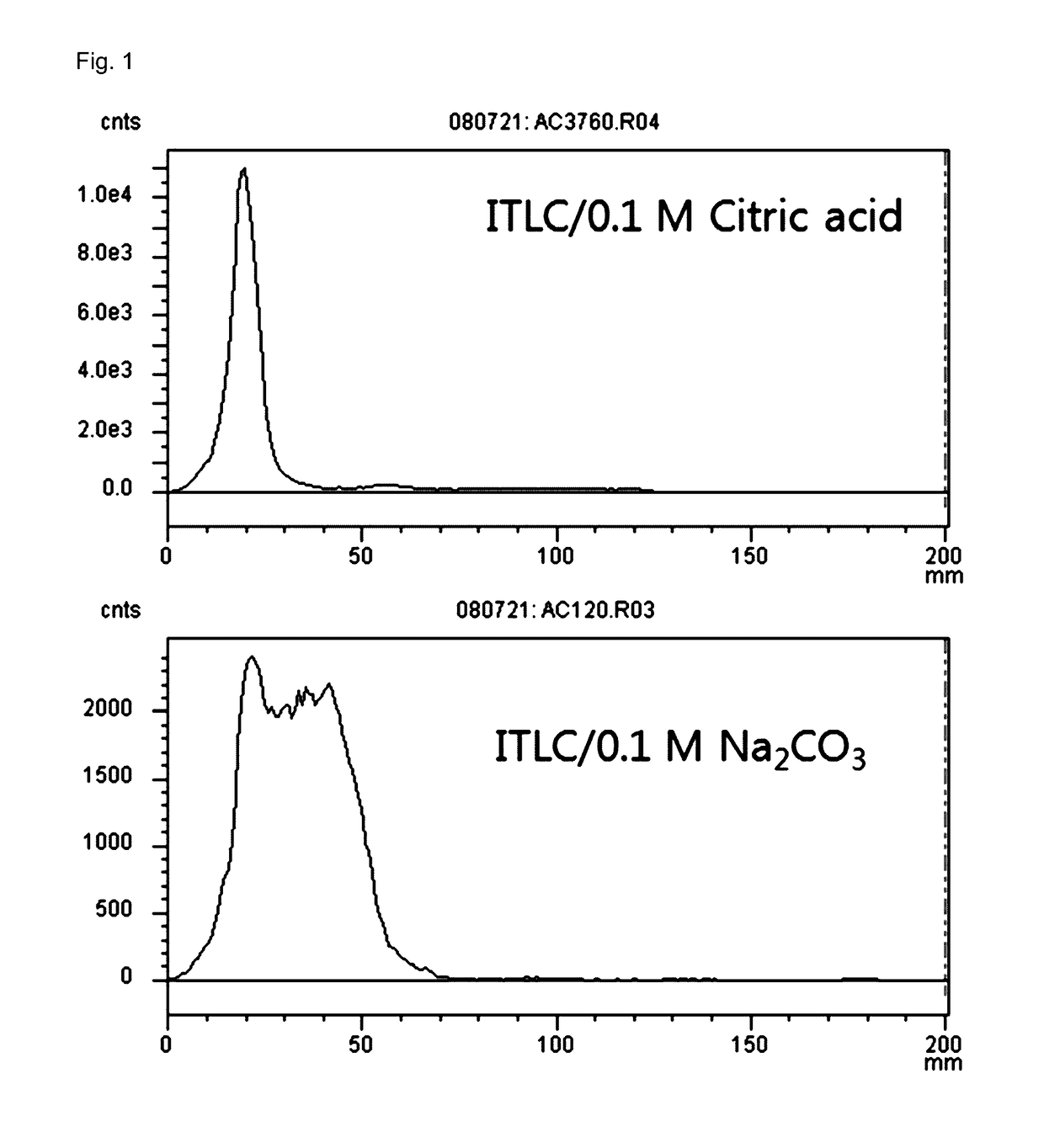
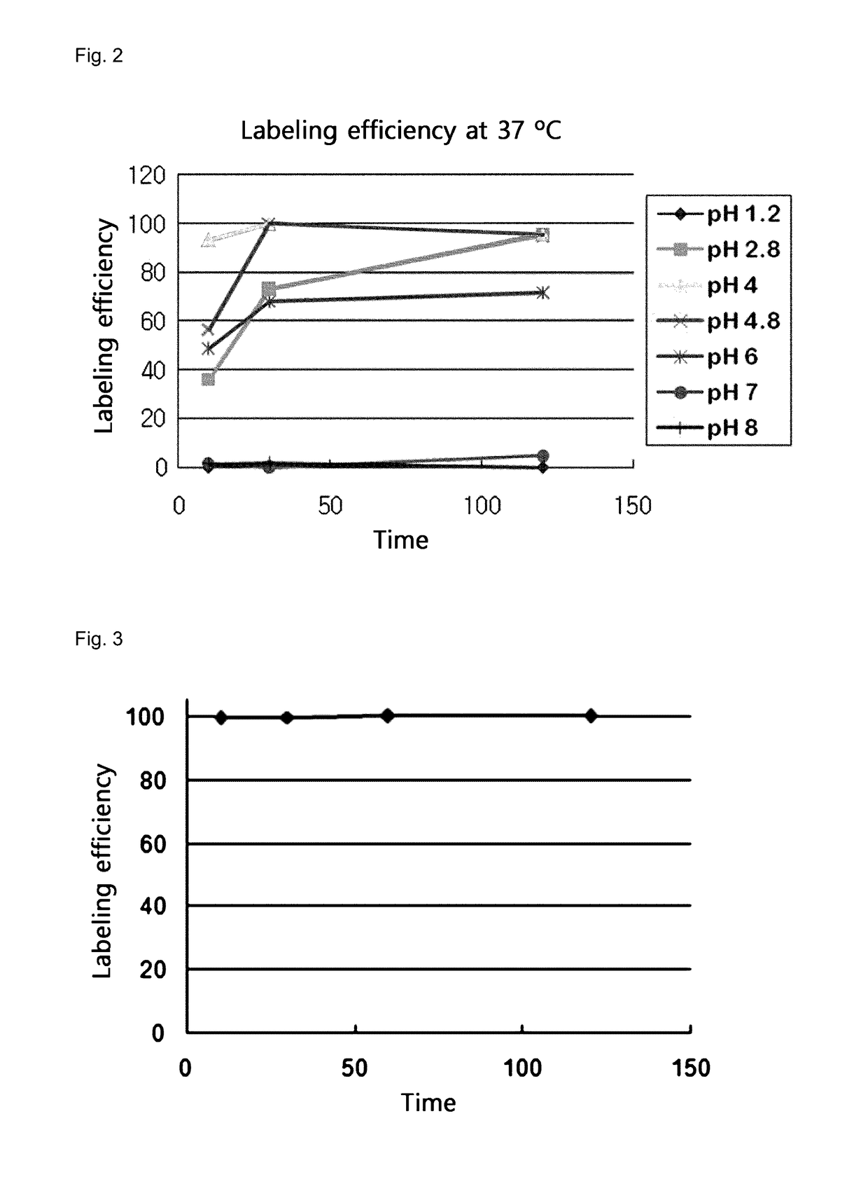
[0034]While conducting reaction at 37° C. after adding 1 mL of a 0.1 M hydrochloric solution of 68GaCl prepared using a 68Ge / 68Ga generator (Cyclotron Co., Russia) to the kit of Example 2, labeling efficiency was measured by TLC 10 minutes, 30 minutes, 1 hour and 2 hours later. ITLC-SG (Gelman Co., USA) was used as a stationary phase and a 0.1 M citric acid solution was used as a mobile phase. The distribution of radioactivity on an ITLC plate was measured using a TLC scanner (Bioscan Co.). The labeled 68Ga remained at the origin and unlabeled 68Ga moved to the solvent front (FIG. 1). The labeling was almost completed after reaction at 37° C. at pH 4-5 for 30 minutes (FIG. 2). The stability of the labeled 68Ga-NOTA-MSA was investigated by measuring radiochemical purity after mixing with human serum and incubation at 37° C. The result is shown in FIG. 3. As can be seen from FIG. 3, when the compound was incuba...

example 4

Preparation of RITC-MSA Compound for Fluorescence Imaging of Mannose Receptor

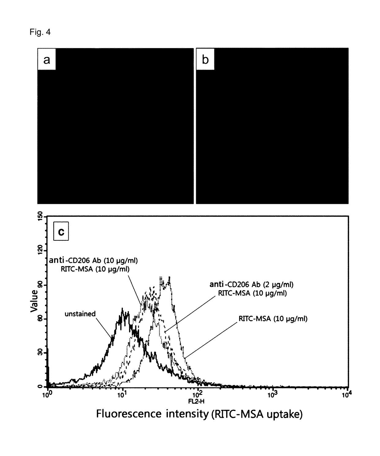
[0035]First, MSA was prepared in the same manner as in the step 1 of Example 1. 100 mg of the MSA was reacted with 16 mg (0.03 mmol) of rhodamine B isothiocyanate (RITC) dissolved in 13 mL of a 0.1 M sodium carbonate buffer (pH 9.5) at room temperature for 20 hours in the dark. The produced RITC-MSA was separated and purified using a PD-10 column and physiological saline and then freeze-dried. The amount of RITC bound per MSA was calculated by measuring molecular weight using a MALDI-TOF mass spectrometer equipped with a nitrogen laser (337 nm). For this, the measurement was made by irradiating laser 500 times in a linear mode. All samples were analyzed 4 times and the molecular weight of MSA and RITC-MSA was determined by averaging the result.

[0036]A composition for imaging atherosclerosis was prepared through tis procedure.

[0037]PET / CT images were obtained for a patient with atherosclerotic symptoms using...


PUM

PropertyMeasurementUnit
Electric chargeaaaaaaaaaa
Capacitanceaaaaaaaaaa
Compositionaaaaaaaaaa

Abstract

The present disclosure relates to a composition for imaging atherosclerosis and a method for diagnosing atherosclerosis using the same. The composition for imaging atherosclerosis according to the present disclosure shows excellent atherosclerosis diagnosis accuracy, enables diagnosis of atherosclerosis even for a person with diseases of glucose metabolism such as diabetes and enables effective diagnosis even for atherosclerosis occurring in the brain and heart. In addition, manufacturing cost is low compared with the existing imaging composition for diagnosis of atherosclerosis. Therefore, atherosclerosis can be effectively diagnosed by using the same.

Description

TECHNICAL FIELD[0001]The present disclosure relates to a composition for imaging atherosclerosis and a method for diagnosing atherosclerosis using the same.BACKGROUND ART[0002]Atherosclerosis refers to a disease in which cholesterol is deposited on the inner portion (intima) of a blood vessel and intimal cells proliferate there, leading to narrowing or blocking of the blood vessel and decreased blood flow to the periphery. More specifically, atherosclerosis is a vascular disease in which, just as an aged water pipe becomes narrower in diameter as rust and impurities are deposited, cholesterol penetrates into intimal layer of a blood vessel and intimal cells and macrophages proliferate there, leading to the formation of an atheroma. The core of the atheroma is composed of acellular lipid core and it is covered by a hard fibrous plaque. When the plaque is unstable, it ruptures and forms blood clots in the blood vessel. Hemorrhage into the atheroma leads to rapid narrowing or even bloc...

Claims


Application Information

Patent Timeline
IPC IPC(8): A61K51/08A61K51/04
CPCA61K51/044A61K51/081A61K51/04A61K51/048A61K51/0482A61K51/0491A61P9/10A61K51/088A61K51/08
Inventor SEO, HONG SEOGKIM, SUNGAURIKIM, EUNG JUJEONG, JAE MIN
Owner KOREA UNIV RES & BUSINESS FOUND
Learn More