Tissue array for cell spheroids and methods of use
a cell spheroids and tissue array technology, applied in the field of tissue arrays for cell spheroids and methods of use, can solve the problems of difficult sectioning and staining several spheroids together, time-consuming and labor-intensive human skeleton analysis, and difficult separation of samples from different groups, so as to save time and money
- Summary
- Abstract
- Description
- Claims
- Application Information
AI Technical Summary
Benefits of technology
Problems solved by technology
Method used
Image

Examples
example 1
Method of Preparing a Microarray of Cell Spheroids.
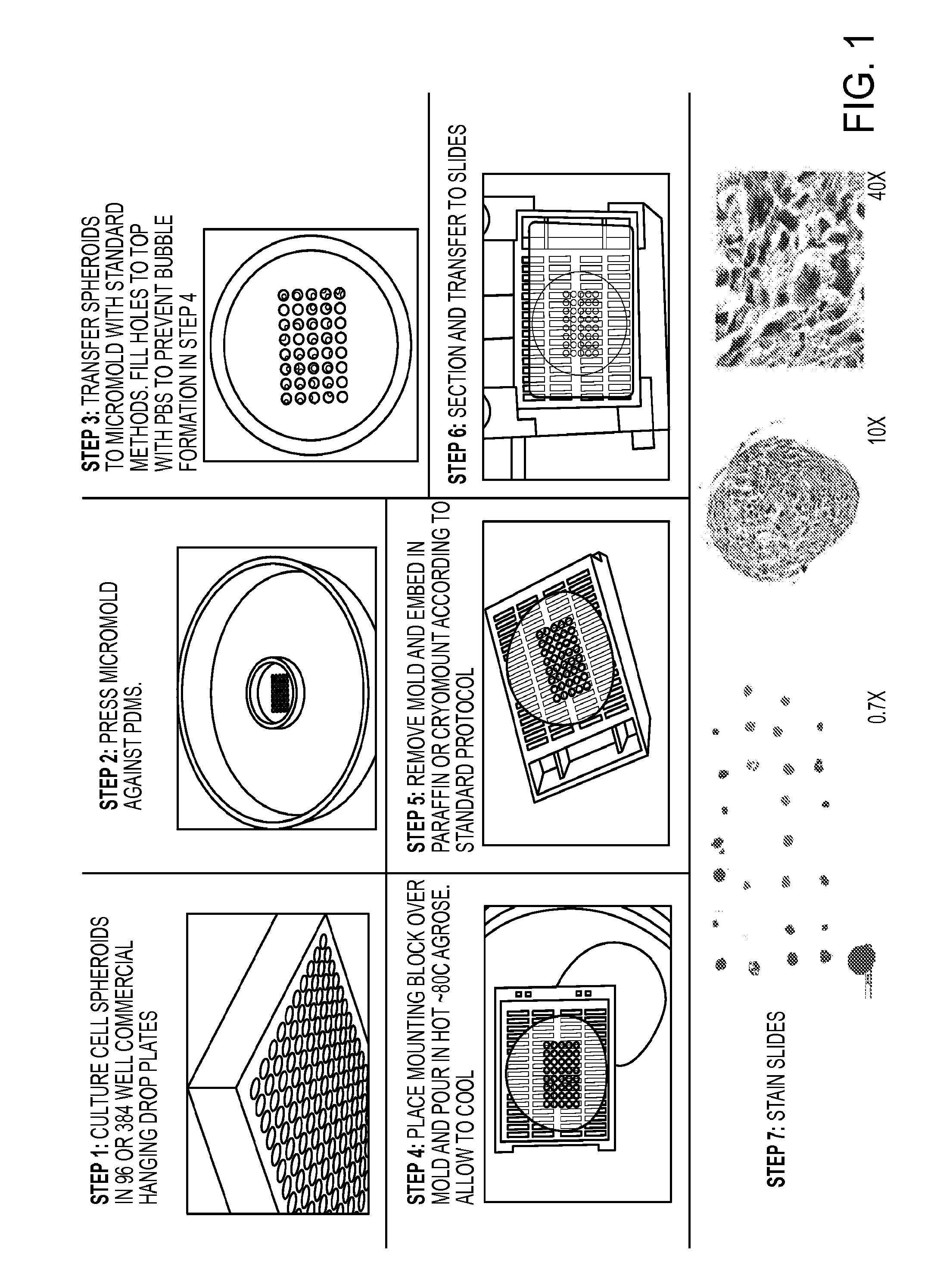
[0098]Histological analysis of cell spheroids is very time consuming if each spheroid is embedded, sectioned, and stained individually. Sectioning and staining several spheroids together is difficult and it becomes especially tricky to keep samples from different groups separated. Here a simple method of sorting and embedding spheroids is presented. This method makes it easy to section many (prototype up to 40) spheroids in the same block and on the same plane, while maintaining the location of each sample. This system is an excellent complement to current advances in scaling up spheroid production in 96 and 384 well plates.
[0099]As a first step, cell spheroids are cultured in in 96 or 384 well commercial hanging drop plates. Next, a micromold is pressed against PDMS. The spheroids are then transferred to the micromold with standard methods, for example with a pipette. Holes are filled to the top with PBS to prevent bubble formation...
experiment 1
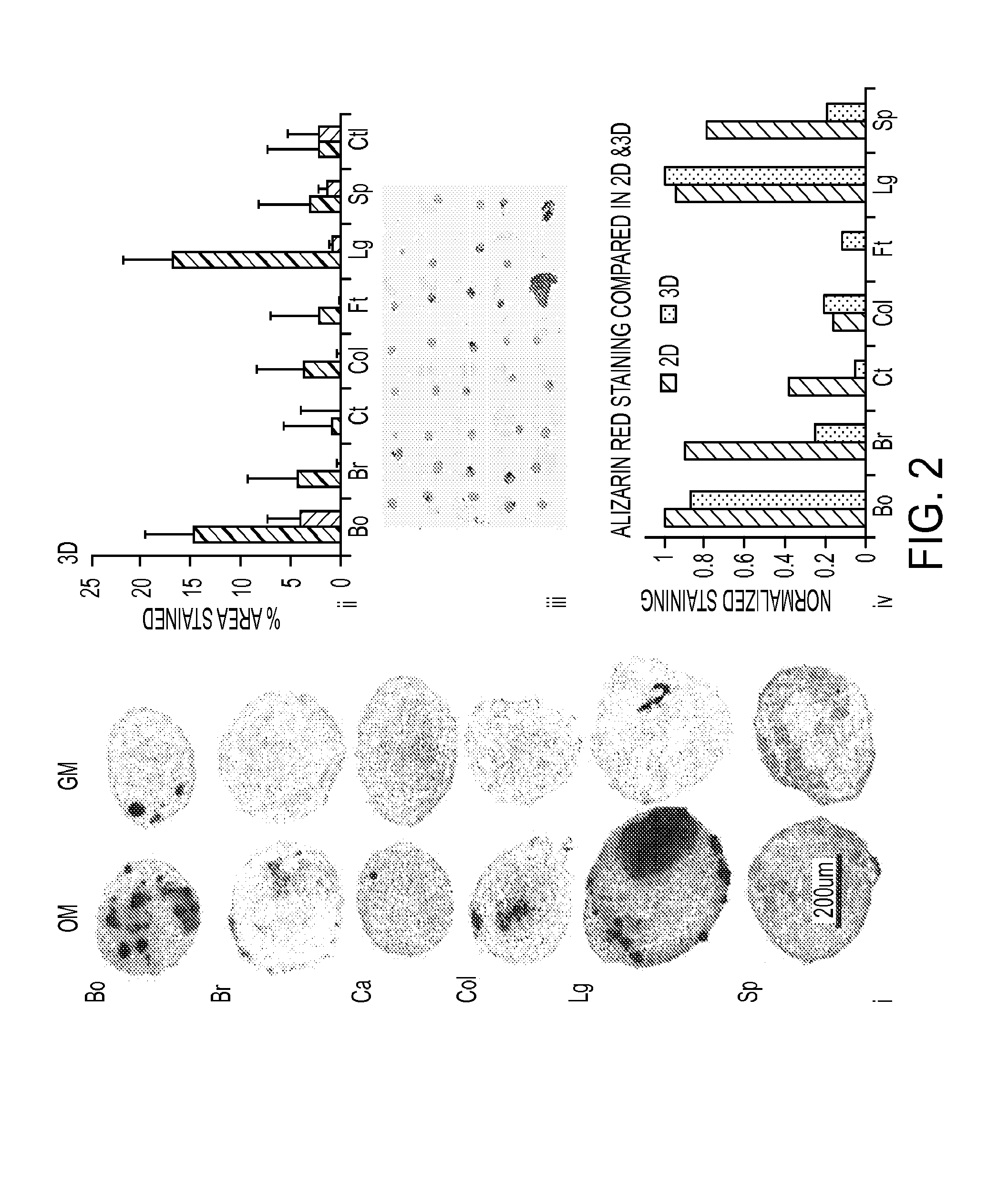
[0115]An experiment was conducted to study the effect of tissue particles on adiopose derived stem cell differentiation in cell / particle spheroids. There were 8 different groups (particle types) and each group was incubated in one of 4 different types of differentiation induction media. To illustrate the novel features of the present invention, if each spheroid was stained and analysed with confocal microscopy each one would have to be stained and imaged individually, and would be limited to 4 total types of stain because of limited channels. If conventional methods of sectioning were used, then 32 separate spheroids would have to be dehydrated, embedded, sectioned, and stained. With the system and methods described herein, all 32 spheroids were able to be sectioned from the microarray and because there were many slides, all 32 spheroids were able to be stained with H&E for cell nuclei and cell / particle organization, Masson's trichrome for extracellular matrix, 2 markers of adipogen...
example 2
Method of Preparing a Microarray of Cell Spheroids Using a Single Piece Micromold.
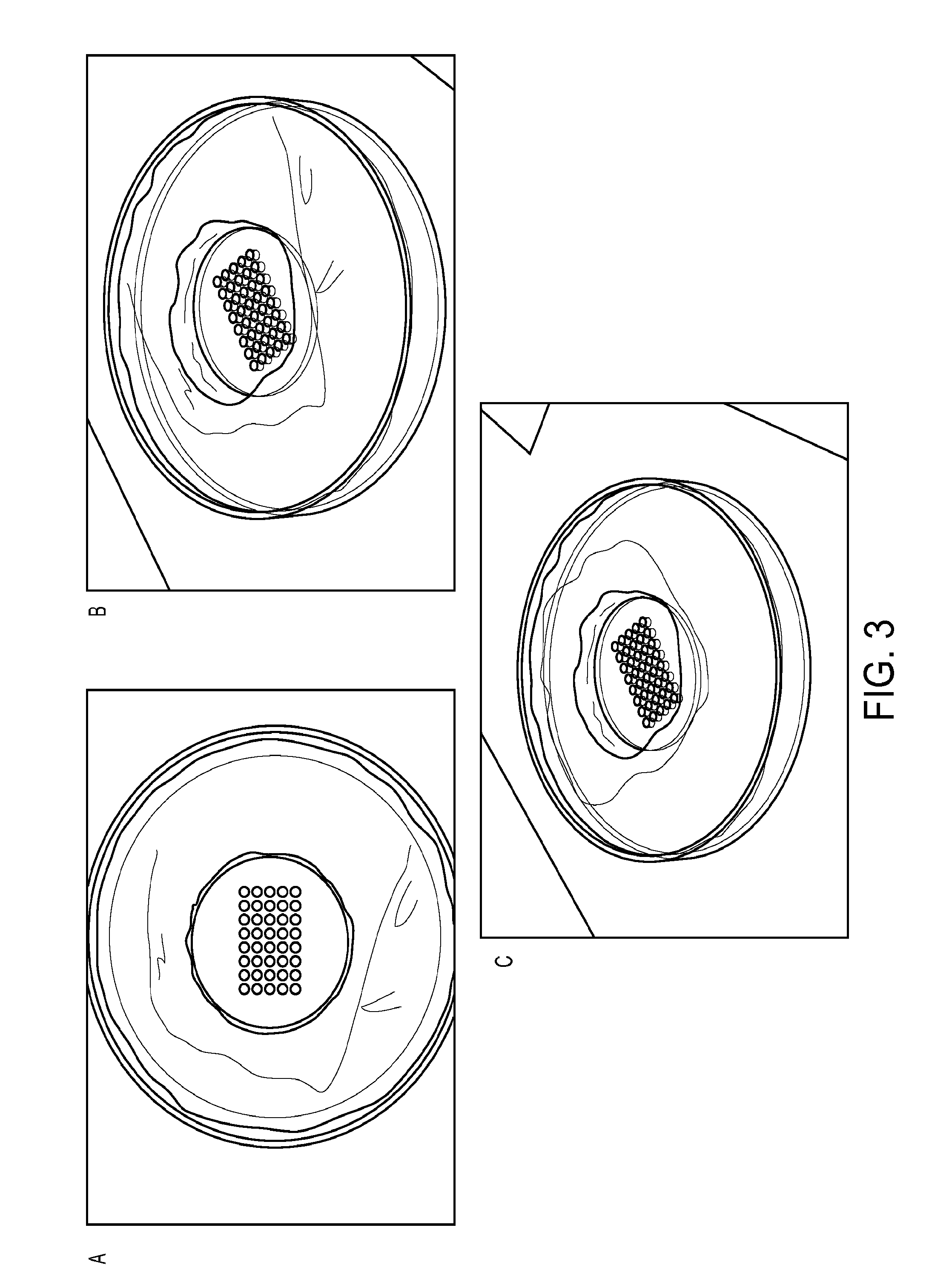
[0117]While certain of the above-exemplified methods for preparing a microarray of cell spheroids involved use of a plastic mold comprising through-holes that was pressed up against a PDMS backing, it was additionally contemplated that a single piece mold could be used in the methods of the invention. A single piece mold comprising an array of wells was therefore synthesized and employed.
[0118]As shown in FIGS. 3A to 3C, a single piece micromold having an array of wells was synthesized from polydimethylsiloxane (PDMS) and was used in the methods of the invention. Testing of the single piece micromold identified it to function as well as the two piece micromold described above (data not shown).
PUM
| Property | Measurement | Unit |
|---|---|---|
| time | aaaaa | aaaaa |
| time | aaaaa | aaaaa |
| time | aaaaa | aaaaa |
Abstract
Description
Claims
Application Information
 - R&D
- Intellectual Property
- Life Sciences
- Materials
- Tech Scout
- Unparalleled Data Quality
- Higher Quality Content
- 60% Fewer Hallucinations
Browse by: Latest US Patents, China's latest patents, Technical Efficacy Thesaurus, Application Domain, Technology Topic, Popular Technical Reports.
© 2025 PatSnap. All rights reserved.Legal|Privacy policy|Modern Slavery Act Transparency Statement|Sitemap|About US| Contact US: help@patsnap.com