
Method for segmentation of the head-neck arteries, brain and skull in medical images

a head-neck artery and medical image technology, applied in image data processing, character and pattern recognition, instruments, etc., can solve the problems of inability to routinely segment large numbers of medical images, material may not be uniformly distributed along the blood vessel, and require a lot of time by skilled professionals

Active Publication Date: 2016-05-05
PHILIPS MEDICAL SYST TECH
View PDF3 Cites 20 Cited by
  • Summary
  • Abstract
  • Description
  • Claims
  • Application Information

AI Technical Summary

Benefits of technology

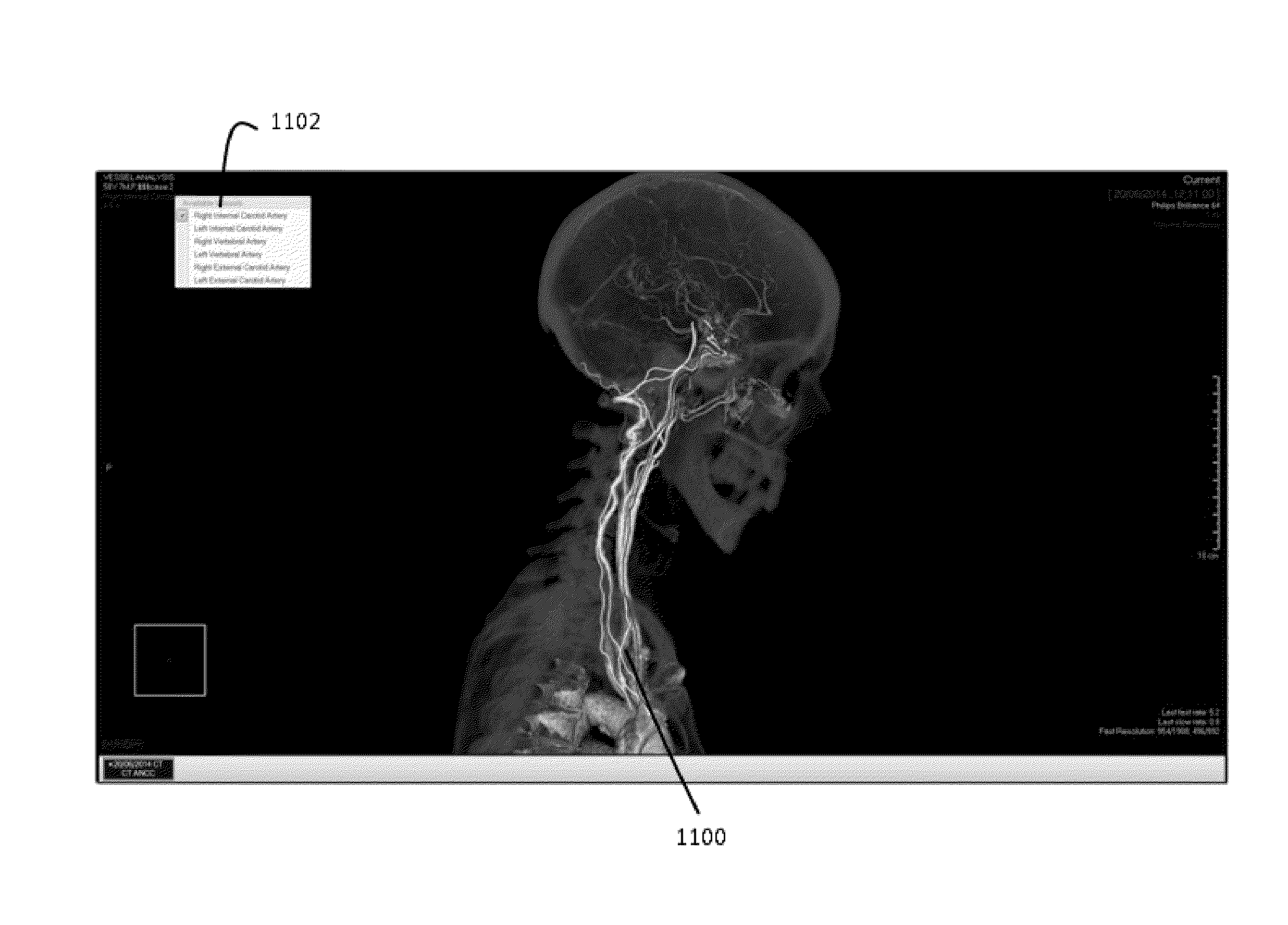
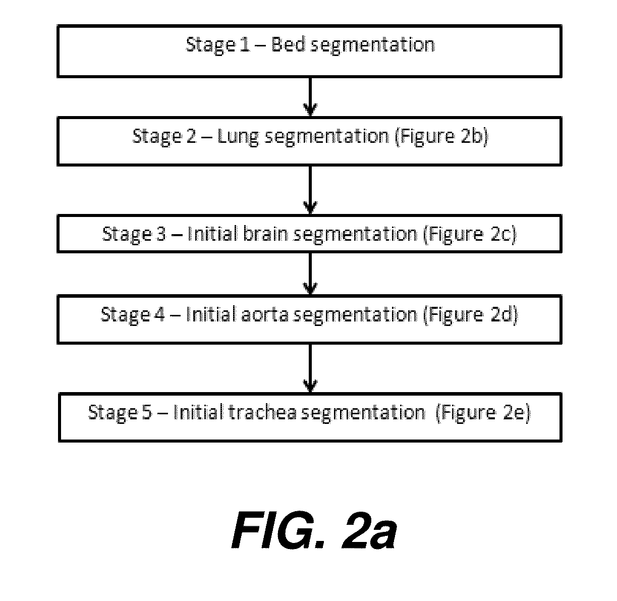
The present invention provides a system and method for automated segmentation of blood vessels in medical images, particularly in the head and neck region. The system and method can identify the location of anatomical landmarks in the image and segment the blood vessels based on the landmarks. This can be useful in identifying blood vessels in medical images, particularly in cases where the blood vessels are not easily visible. The system and method can also identify the starting and ending points of the blood vessels and define a path between them. This can be useful in analyzing blood flow in the brain and skull. The method can be performed semi-automatically, with the user only identifying the landmarks and the relevant landmark slices. Overall, the system and method provide a more efficient and accurate way to segment blood vessels in medical images.

Problems solved by technology

a) Even if the blood vessel is imaged using an intravenous radiocontrast agent, this material may not be uniformly distributed along the blood vessel;
b) Partial volume effects, especially near bones;
c) Nearby tissue with similar absorption, mainly various bones that have similar HU values;
d) Narrowing, splitting and / or other geometrical properties of vessels;
e) Nearby vessels may appear to meet and merge and then diverge;
Prior art segmentation tools have generally required at least some manual user input, such as identifying the starting point and ending points of the neck arteries, and segmenting each image can therefore require a lot of time by skilled professionals, making it impractical to routinely segment large numbers of medical images.

Method used


Image

  • Method for segmentation of the head-neck arteries, brain and skull in medical images
  • Method for segmentation of the head-neck arteries, brain and skull in medical images
  • Method for segmentation of the head-neck arteries, brain and skull in medical images

Examples


Embodiment Construction

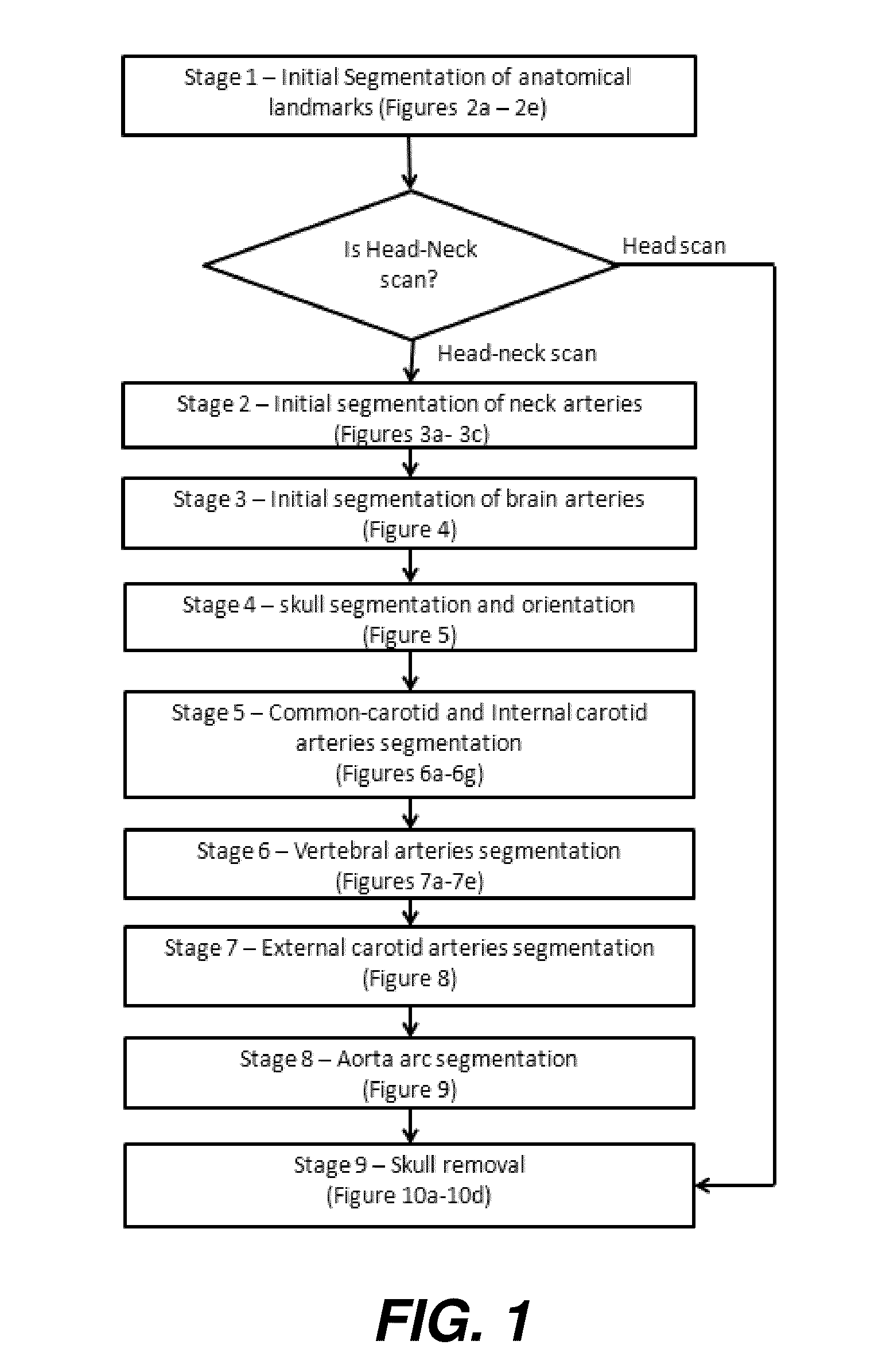
[0067]The present invention provides a system and method, in at least some embodiments, for manipulation of head-neck CT scan images to automatically segment the brain and the head-neck arteries preferably including the main head-neck arteries: the common, internal and external carotid arteries, the vertebral arteries on the left and right side, the aortic arch, and the basilar artery. Optionally, a user aided recovery process is provided in case the automated method fails that allows the automated method to continue after some manual input. The algorithms and methods described are optimized to enable automated segmentation in a minimum amount of time.

[0068]The resulting segmentation preferably enables display and labeling on a medical image viewer of the head-neck arteries and further preferably enables removal from the displayed image of the brain, skull, vertebral bones and all other bones in the image along with any other non-brain soft tissue, air cavities and any other parts o...


PUM


Abstract

A method for automated segmentation of a blood vessel of a head and neck of a subject in a medical image, the method comprising: identifying the location of anatomical landmarks in the medical image; identifying regions of interest in the medical image based on the landmarks; segmenting segments of blood vessels in the medical image; classifying at least one of the segments as defining the blood vessel based on its position relative to the landmarks within the regions of interest to create a classified blood vessel; identifying a starting seed for the blood vessel from the classified blood vessel; identifying an ending seed for the blood vessel from the classified blood vessel; segmenting the blood vessel between the starting seed and the ending seed; and defining a path between the starting seed and the ending seed.

Description

CROSS REFERENCE TO RELATED APPLICATIONS[0001]This application claims priority to U.S. Ser. No. 62 / 074,223 provisionally filed on Nov. 3, 2014, titled SYSTEM AND METHOD FOR SEGMENTATION OF MEDICAL IMAGES, to Silbert et al, incorporated herein in its entirety.FIELD OF THE INVENTION[0002]The present disclosure relates to a system and method for medical image manipulation and in particular, such a system and method for segmenting the brain, the skull and segmentation of the head-neck arteries in medical images.BACKGROUND OF THE INVENTION[0003]CT (computed tomography) imaging systems can be used by radiologists to examine the blood vessels of the head and neck including degree of stenosis (narrowing) or aneurisms. Radiologists analyze the vessels in two scenarios. In one scenario each vessel is examined separately, traversing along the vessel and checking for aneurisms or stenosis. The second method is by using a MIP (Maximum Intensity Projection) view of the head and examining the overa...

Claims


Application Information

Patent Timeline
Patent Type & Authority Applications(United States)
IPC IPC(8): G06T7/00G06T11/00
CPCG06T7/0012G06T11/003G06T2207/30061G06T2207/30101G06T2207/30008G06T2207/10081G06T2207/20036G06T2207/20156G06T7/11
Inventor SILBERT, OHADSHREIBER, REUVEN R.ENGELHARD, GUY E.PORAT, HADARGAZIT, TIFERET T.
Owner PHILIPS MEDICAL SYST TECH
Learn More