
Method for computationally predicting optimal placement sites for internal defibrillators in pediatric and congenital heart defect patients

a technology of congenital heart defect and optimal placement site, which is applied in the field of computational model for determining the placement of cardiac cardioverter/defibrillator devices, can solve the problems of pain and psychological trauma, no reliable, personalized prediction method, and use of non-standard icd configurations, and achieve low defibrillation. the effect of removing breathing motion errors

Active Publication Date: 2016-04-28
THE JOHN HOPKINS UNIV SCHOOL OF MEDICINE
View PDF2 Cites 18 Cited by
  • Summary
  • Abstract
  • Description
  • Claims
  • Application Information

AI Technical Summary

Benefits of technology

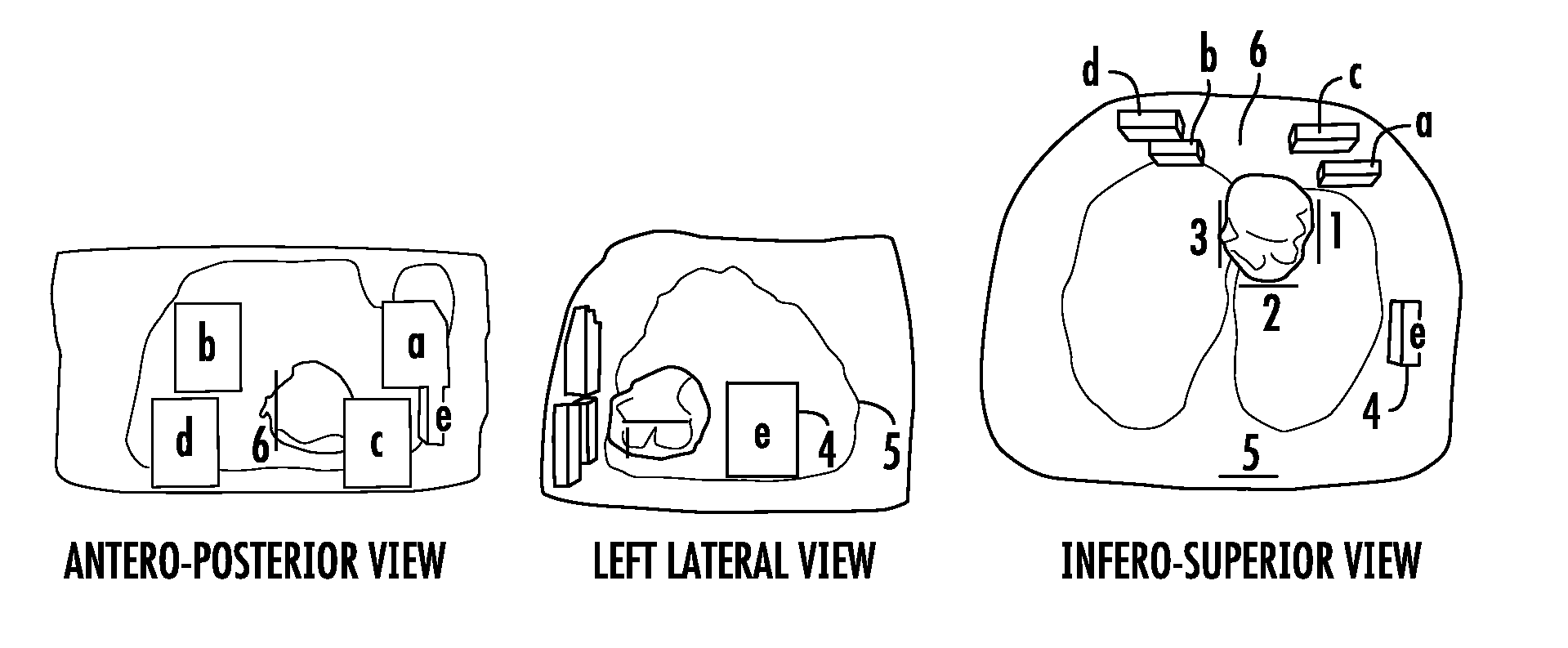
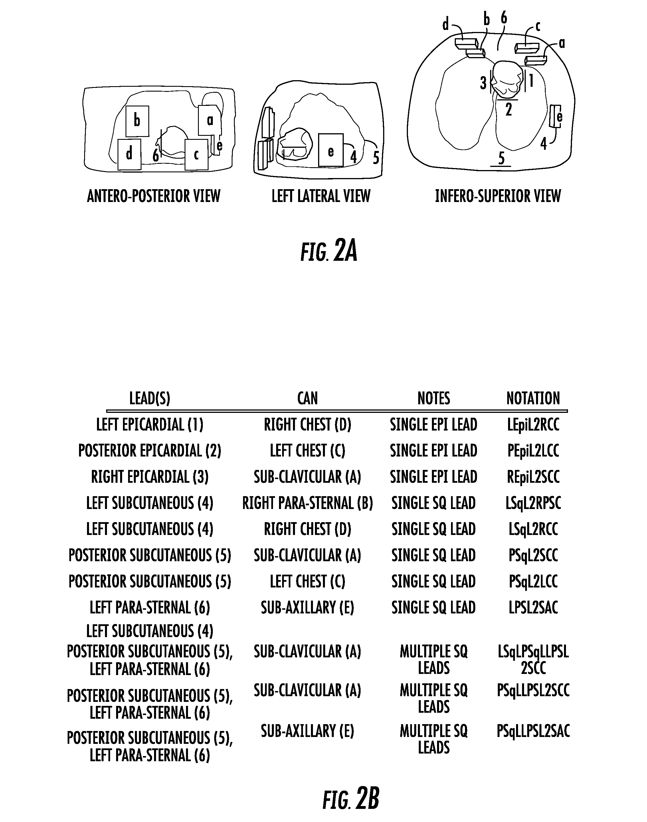
The present invention provides a method for creating a magnetic resonance image of a ventricular model that can help in the placement of a cardiac defibrillator. The method includes assigning fiber and sheet orientations to the ventricular model, creating a binary image of ventricular geometry, and eliminating breathing motion errors. The method can also include adding an area of interest to the model, based on the bidomain and monodomain representations of current flow in cardiac tissue. The method can help in choosing the cardiac defibrillator placement with the lowest defibrillation and cardioversion thresholds or the placement that is otherwise deemed favorable by the responsible health care provider.

Problems solved by technology

In these patients, it is often not indicated or even impossible to implant a transvenous lead ICD because of the patient's small heart size and congenitally altered anatomy, thus non-standard ICD configurations have to be used.
However, there is currently no reliable, personalized way of predicting which ICD configuration would have the lowest defibrillation threshold (DFT) and cardioversion threshold (CVT) in a patient.
A low DFT is desirable because strong electric shocks damage cardiac myocytes, increase mortality, and can cause pain and psychological trauma.

Method used


Image

  • Method for computationally predicting optimal placement sites for internal defibrillators in pediatric and  congenital heart defect patients
  • Method for computationally predicting optimal placement sites for internal defibrillators in pediatric and  congenital heart defect patients
  • Method for computationally predicting optimal placement sites for internal defibrillators in pediatric and  congenital heart defect patients

Examples


example

[0030]An exemplary implementation of the present invention is described herein, in order to further illustrate the present invention. The exemplary implementation is included merely as an example and is not meant to be considered limiting, Any implementation of the present invention on any suitable subject known to or conceivable by one of skill in the art could also be used, and is considered within the scope of this application.

Data Acquisition

[0031]A clinical MRI (1.5 T) dataset acquired from a 14-year-old male pediatric patient with tricuspid valve atresia is used throughout this example. Because of the tricuspid valve atresia, the right ventricle (RV) was not recognizable on the clinical low-resolution MRI images. This dataset included axial torso scans, and short-axis (SA) and long-axis (LA) cardiac cine scans. The axial image (27 slices, 384×300 pixels each) had a resolution of 1×1×6 mm3, and the SA cine scans (11 slices, 144×192 pixels each) had a resolution of 2×2×10 mm3, a...


PUM


Abstract

The present invention includes a method for determining optimal placement sites for internal defibrillators in pediatric and congenital heart defect patients. The method is executed by creating a personalized active heart-torso model. The model is created using imaging scans (e.g., low resolution clinical scans) and advanced image processing techniques. The image processing results in a heart-torso mesh model. The ventricular portion of the mesh incorporates cell membrane dynamics. The combined torso-active ventricular defibrillation model can be used for patient specific modeling of the defibrillation process and optimal defibrillator placement can be determined. This method could also be used to decrease the energy needed for a defibrillation shock, because of the optimized defibrillator placement.

Description

CROSS REFERENCE TO RELATED APPLICATIONS[0001]This application claims the benefit of U.S. Provisional Patent Application 61 / 820,974 filed on May 8, 2013, which is incorporated by reference, herein, in its entirety.GOVERNMENT SUPPORT[0002]This invention was made with government support under R01 HL103428 awarded by the National Institutes of Health. The government has certain rights in the invention.FIELD OF THE INVENTION[0003]The present invention relates generally to cardiology. More particularly the present invention relates to a computational model for determining placement of cardiac cardioverter / defibrillator devices.BACKGROUND OF THE INVENTION[0004]Defibrillation by strong electric shock is the only known procedure that reliably terminates ventricular fibrillation (VF). Implantable cardioverter delibrillators (ICDs) have recently been implanted with increasing frequency in the pediatric population and in patients with congenital heart defects (CHD). In these patients, it is oft...

Claims


Application Information

Patent Timeline
Patent Type & Authority Applications(United States)
IPC IPC(8): A61B34/10A61N1/39
CPCA61B34/10A61B2034/105A61B2034/104A61N1/3956A61N1/3918
Inventor TRAYANOVA, NATALIARANTNER, LUKASVADAKKUMPADAN, FIJOY
Owner THE JOHN HOPKINS UNIV SCHOOL OF MEDICINE
Learn More