
Ultrasonic diagnostic apparatus and method thereof

a diagnostic apparatus and ultrasonic technology, applied in ultrasonic/sonic/infrasonic image/data processing, ultrasonic/sonic/infrasonic diagnostics, tomography, etc., can solve problems such as difficulty in accurately calculating three-dimensional blood flow vector information

Inactive Publication Date: 2015-12-24
INST FOR BASIC SCI
View PDF2 Cites 3 Cited by
  • Summary
  • Abstract
  • Description
  • Claims
  • Application Information

AI Technical Summary

Benefits of technology

The present invention provides an ultrasonic diagnostic apparatus and method that can accurately calculate the velocity of blood flow in the heart or other organs without injecting contrast enhancing media. This is done by extracting data from 2D B-mode echocardiograms and Doppler images, using a 2D Navier-Stokes equation with a mass source term. The method can be implemented using existing Doppler data for C-mode images, without changing hardware design. This allows for improved and non-invasive diagnosis of cardiovascular disease.

Problems solved by technology

However, such a technique has disadvantages in that contrast enhancing media are necessarily injected to calculate information on vector components of the blood flow within the heart, and capabilities of tracing speckles of the blood flow seriously depend upon image quality.
For this reason, it is difficult to accurately calculate three-dimensional blood flow vector information.

Method used


Image

  • Ultrasonic diagnostic apparatus and method thereof
  • Ultrasonic diagnostic apparatus and method thereof
  • Ultrasonic diagnostic apparatus and method thereof

Examples


Embodiment Construction

[0029]Hereinafter, exemplary embodiments of the present invention will be described in detail with reference to the accompanying drawings. The following embodiments are given by way of illustration to provide a thorough understanding of the disclosure to those skilled in the art. It should be understood that the present invention is not limited to the following embodiments and may be embodied in different ways. In the drawings, portions irrelevant to the description will be omitted for clarity. Like components will be denoted by like reference numerals throughout the specification.

[0030]It will be understood that the terms “comprise,”“include,” and / or “have(has)” as used in this specification, specify the presence of stated features, steps, operations, elements, and / or components, but do not preclude the presence or addition of one or more other features, steps, operations, elements, components, and / or groups.

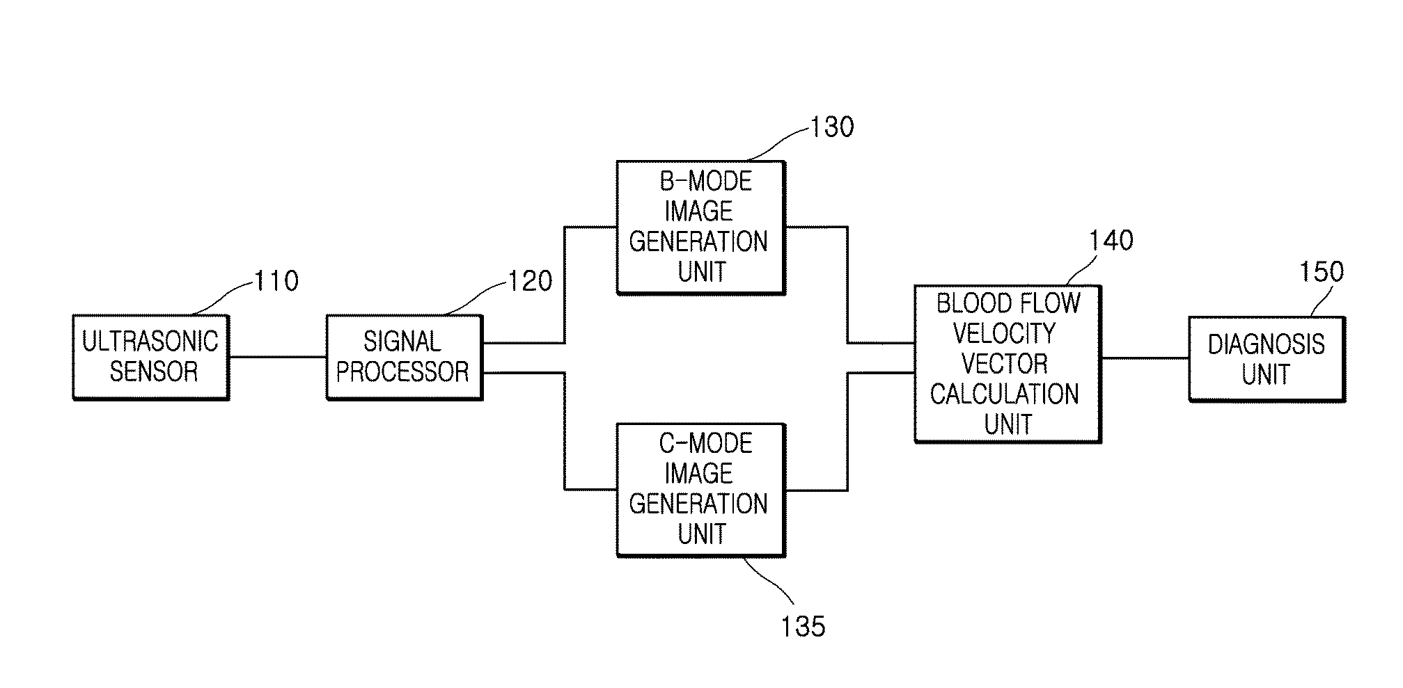
[0031]FIG. 1 is a view showing features of an ultrasonic diagnostic appara...


PUM


Abstract

Disclosed is an ultrasonic diagnostic apparatus. The ultrasonic diagnostic apparatus includes: an ultrasonic sensor transmitting ultrasonic waves and sensing echo signals thereof; a signal processor processing the signals sensed by the ultrasonic sensor; a B-mode image generation unit generating a brightness mode (B-mode) image in a 2D plane based on the signal processing results by the signal processor and extracting left ventricle boundary data from the generated B-mode image; a C-mode image generation unit generating Doppler data for generation of a C-mode image based on the signal processing results by the signal processor; and a blood flow velocity vector calculation unit calculating a blood flow velocity vector in a 2D image plane based on a simultaneous equation composed of a relation equation between the extracted left ventricle boundary data, the Doppler data and the blood flow velocity vector, and a 2D Navier-Stokes equation obtained from image data generated through signal processing by the signal processor, wherein the Navier-Stokes equation further uses, as a variable, a mass source term representing distribution of source and sink of mass with respect to the 2D image plane.

Description

BACKGROUND[0001]1. Technical Field[0002]The present invention relates to an ultrasonic diagnostic apparatus and an ultrasonic diagnostic method thereof, and more particularly, to an ultrasonic diagnostic apparatus, which can exactly calculate a velocity vector of a blood flow in the heart or the like using left ventricle boundary data extracted from 2D brightness mode (B-mode) echocardiograms and Doppler data for generation of color Doppler mode (C-mode) images, without injection of contrast agents (or contrast enhancing media) into blood vessels in ultrasonic diagnosis of the heart or the like, and an ultrasonic diagnostic method thereof.[0003]2. Description of the Related Art[0004]Ultrasonic diagnostic apparatuses are widely used in medicine to acquire information on an internal structure of a subject due to their noninvasive and nondestructive characteristics. Further, ultrasonic diagnostic apparatuses are essentially used in medicine by virtue of their capabilities of providing ...

Claims


Application Information

Patent Timeline
Patent Type & Authority Applications(United States)
IPC IPC(8): A61B8/06A61B8/08A61B8/14
CPCA61B8/06A61B8/14A61B8/0883A61B8/5207A61B8/5223A61B8/488A61B8/0891A61B8/5246G16H50/30A61B8/08
Inventor AHN, CHI YOUNGJEON, KI WANCHOI, JUNG II
Owner INST FOR BASIC SCI
Learn More