
System, method, and computer-accessible medium for determining at least one characteristic of at least one tissue or at least one MRI measurement condition of the at least one tissue using active contrast encoding magnetic resonance imaging procedure(s)

a technology of magnetic resonance imaging and at least one characteristic, applied in the field of magnetic resonance imaging, can solve the problems of limited diagnostic accuracy, further increase of scan time, and difficulty in quantitative analysis of dce-mri data, and achieve the effect of enhancing the contrast of the magnetic resonance imaging curv

Active Publication Date: 2015-10-29
NEW YORK UNIV
View PDF4 Cites 10 Cited by
  • Summary
  • Abstract
  • Description
  • Claims
  • Application Information

AI Technical Summary

Benefits of technology

The patent describes a system, method, and computer-accessible medium for generating images of tissue using magnetic resonance imaging. The system receives magnetic resonance information about the tissue, including the intensity of signals from a pulse sequence. The system uses flip angles and a pulse sequence to encode the signals and determine the tissue characteristics, such as proton density and relaxation time. The system can also account for imperfections in the imaging process, such as B1 inhomogeneity and nonlinearity of the radio frequency amplifier. The system can be used to determine the vascular micro-environmental properties of the tissue. The generated images can provide valuable information for diagnostic purposes.

Problems solved by technology

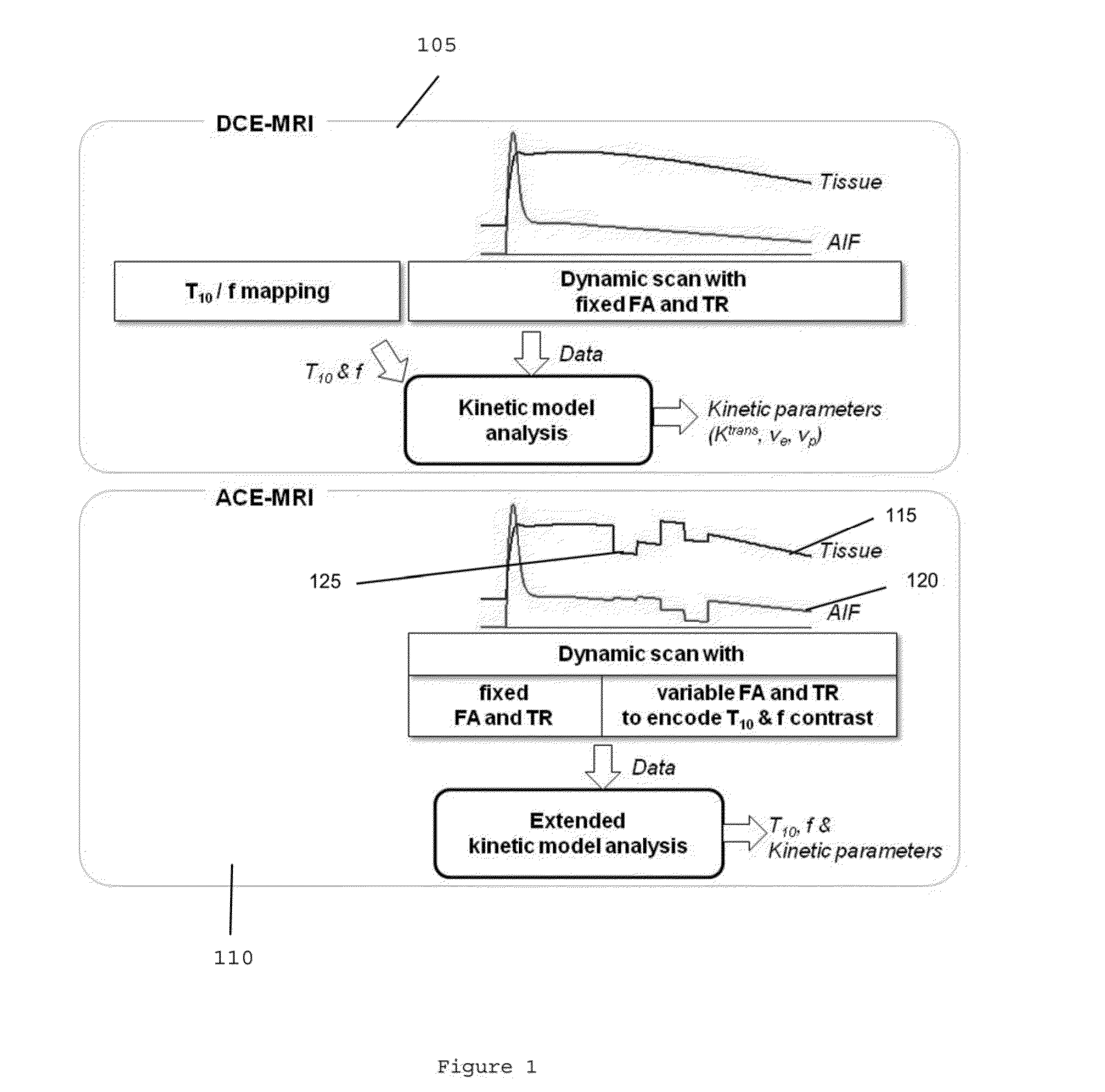
However, quantitative analysis of DCE-MRI data remains challenging, particularly due to the need to separately measure the pre-contrast longitudinal relaxation time constant (e.g., T10) of the tissue (see, e.g., Reference 9) and actual flip-angles (“FA”) achieved for T1 measurement and the dynamic scan.
The degree of contrast enhancement in a tissue can vary depending on the T10 of the tissue, such that analyses of DCE-MRI data based on time-intensity curves without taking into account the T10 variability in lesions can result in a limited diagnostic accuracy.
Most B1 mapping methods utilize either a long repetition time (“TR”) to minimize the tissue T1 effect, or an extra measurement of B0 field to minimize the off-resonance effect, which can lead to a further increase of the scan time.
Since the image magnitude can depend on T1 of tissue and TR, the magnitude-based methods typically utilize a long TR (e.g., >about 5T1) to eliminate the T1 dependence (see, e.g., Reference 21), which can lead to a long scan time.
One of the challenging issues with the phase-based methods can be the influence from the B0 inhomogeneity that can bring a need for additional B0 field mapping at the cost of additional scan time, or careful advanced design of RF pulse.
Thus, while these recent developments offer a number of different ways to measure B1 field map, acquiring B1 map during clinical scans still remains technically challenging and an extra burden of scan time and warrants further development.

Method used


Image

  • System, method, and computer-accessible medium for determining at least one characteristic of at least one tissue or at least one MRI measurement condition of the at least one tissue using active contrast encoding magnetic resonance imaging procedure(s)
  • System, method, and computer-accessible medium for determining at least one characteristic of at least one tissue or at least one MRI measurement condition of the at least one tissue using active contrast encoding magnetic resonance imaging procedure(s)
  • System, method, and computer-accessible medium for determining at least one characteristic of at least one tissue or at least one MRI measurement condition of the at least one tissue using active contrast encoding magnetic resonance imaging procedure(s)

Examples


Embodiment Construction

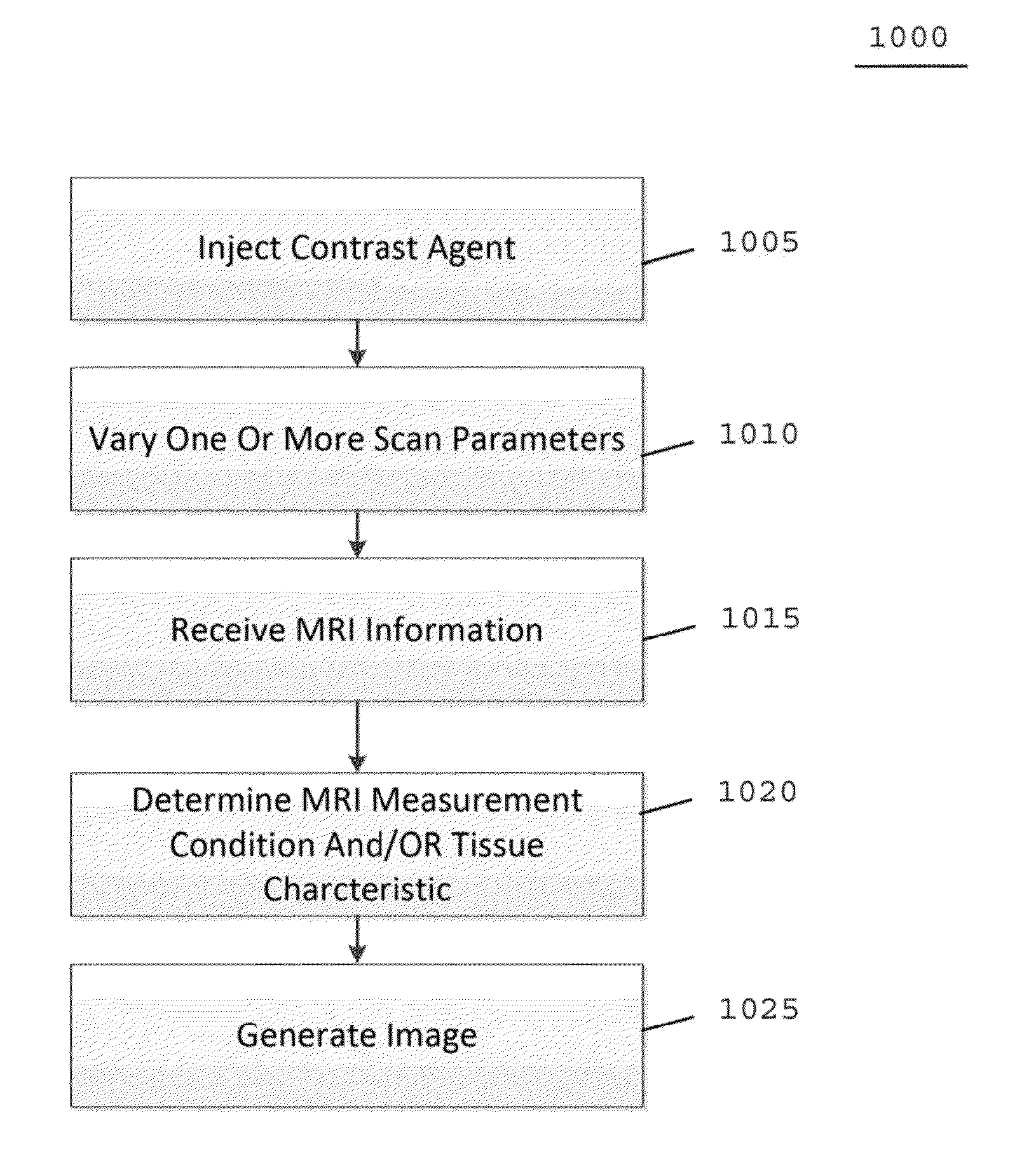
[0008]An exemplary system, method and computer-accessible medium for generating an image of a tissue(s) can be provided, which can include, for example, receiving magnetic resonance imaging information regarding the tissue(s) including an intensity(s) of a signal(s) provided from the tissue(s), actively encoding the signal intensity(s) with a flip angle(s) and a repetition time(s) of the pulse sequence (s), and generating the image of the tissue(s) based on the encoded signal intensity(s). A flip angle correction factor(s), and a T10 value(s), can be embedded in the magnetic resonance information. The flip angle correction factor(s) can be based on a (i) B1 field inhomogeneity, (ii) a radio frequency pulse profile, (iii) a nonlinearity of a radio frequency amplifier, (iv) properties of the tissue(s), and / or (v) imperfect spoiling in steady-state imaging.

[0009]In some exemplary embodiments of the present disclosure, the flip angle(s) can be scaled based on the flip angle correction f...


PUM


Abstract

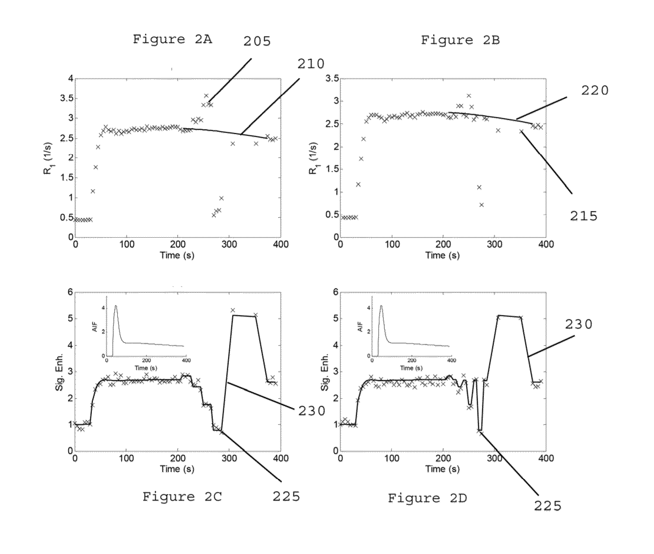
In another exemplary embodiment of the present disclosure is an exemplary system, method and computer-accessible for determining a characteristic(s) of a tissue(s), that can include, for example, receiving magnetic resonance imaging information regarding the tissue(s) including a time-intensity curve(s) of the tissue(s) based on a contrast agent(s) concentration, actively encoding a part of the time-intensity curve(s) with a magnetic resonance relaxation property(s) of the tissue(s) by varying a magnetic resonance imaging scan parameter(s) to generate encoded data during magnetic resonance data acquisition, and determining the tissue characteristic(s) based on the encoded data.

Description

CROSS-REFERENCE TO RELATED APPLICATION(S)[0001]This application relates to and claims priority from U.S. Patent Application No. 61 / 984,384, filed on Apr. 25, 2014, the entire disclosure of which is incorporated herein by reference.STATEMENT REGARDING FEDERALLY SPONSORED RESEARCH[0002]This invention was made with government support under Grant No R01CA160620 awarded by the National Institute of Health. The government has certain rights in the invention.FIELD OF THE DISCLOSURE[0003]The present disclosure relates generally to magnetic resonance imaging (“MRI”), and more specifically, to exemplary embodiments of systems, methods, and computer-accessible mediums for determining at least one characteristic of at least one tissue or at least one MRI measurement condition of the at least one tissue, by measuring, for example, pharmacokinetic model parameters, pre-contrast magnetic resonance (“MR”) relaxation properties (e.g., T10 and T20*) and MR measurement conditions (e.g. B1 and B0) usin...

Claims


Application Information

Patent Timeline
IPC IPC(8): G01R33/56A61B5/055G01R33/32
CPCG01R33/5601A61B2576/00A61B5/055G01R33/32G01R33/24G01R33/243G01R33/246G01R33/561G01R33/56366G01R33/56563G01R33/5659
Inventor KIM, SUNGHEON GENEZHANG, JIN
Owner NEW YORK UNIV
Learn More