Artificial skin tissue, artificial skin model and manufacturing method therefor
a technology of artificial skin and skin tissue, applied in the field of artificial skin tissue and artificial skin models, can solve the problem of animal welfare consideration of animal experiments
- Summary
- Abstract
- Description
- Claims
- Application Information
AI Technical Summary
Benefits of technology
Problems solved by technology
Method used
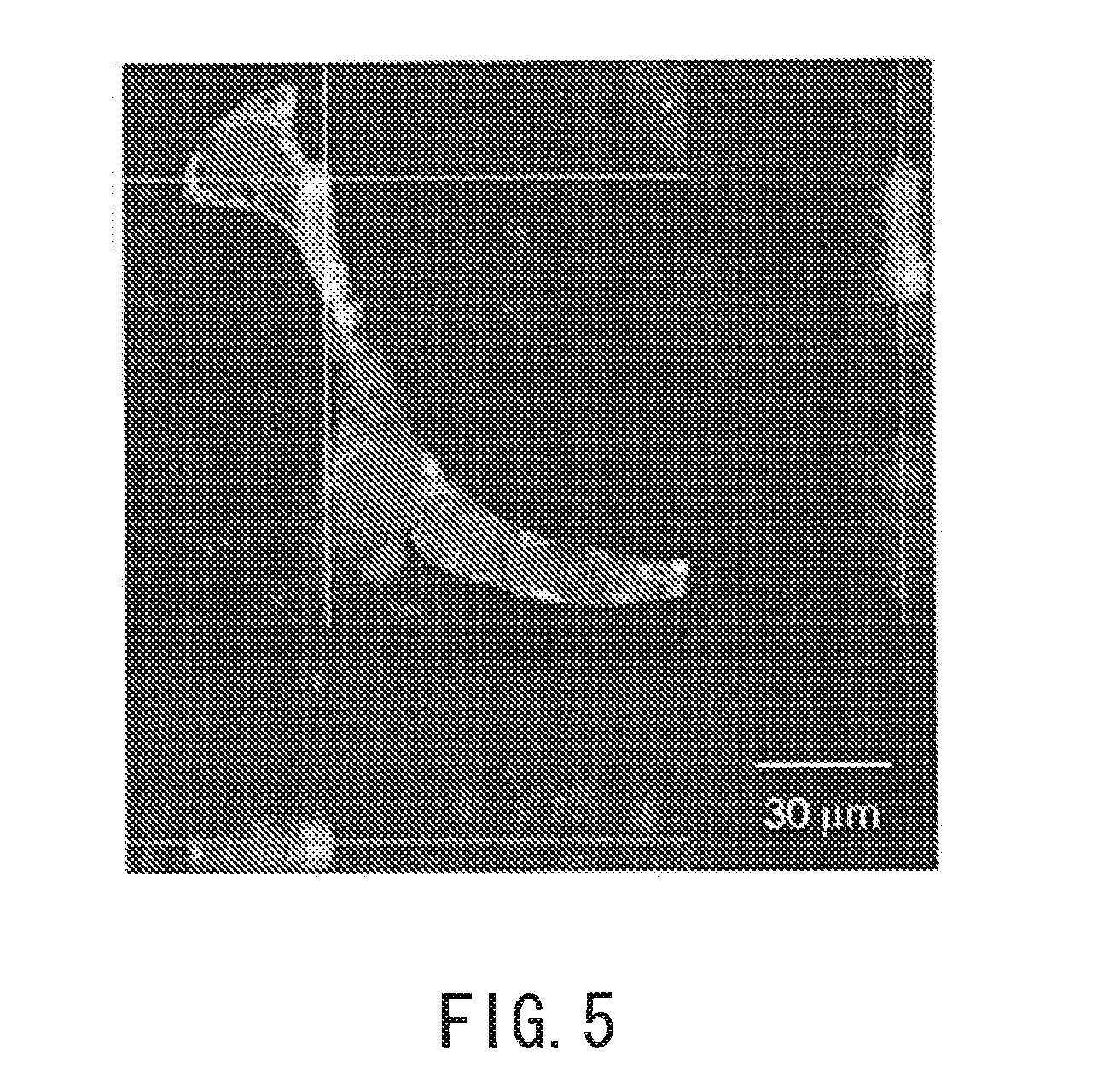
Image


Examples
example 1
Preparation of Coated Cells
[0132]Human fibroblasts (normal human dermal fibroblasts (NHDF), produced by Cambrex Corporation) were dispersed at a concentration of 1×10⁶ cell / ml in a 50 mM Tris-HCl solution (pH=7.4) containing 0.2 mg / ml fibronectin. The dispersion state was maintained for 1 minute while this solution was gently stirred by inverting the container. Then, the solution was centrifuged at 2500 rpm for 1 minute (FN immersion operation). After the supernatant was removed, a 50 mM Tris-HCl solution (pH=7.4) was added so that the cells were dispersed. The dispersion state was maintained for 1 minute while this solution was gently stirred by inverting the container. Then, the solution was centrifuged at 2500 rpm for 1 minute (washing operation). After the supernatant was removed, the cells were dispersed in a 50 mM Tris-HCl solution (pH=7.4) containing 0.2 mg / ml gelatin. The dispersion state was maintained for 1 minute while this solution was gently stirred by inverting the con...
example 2
[0140]An artificial skin model was prepared in the same manner as Example 1 except that 5×10⁴ MoDC coated cells and 5×10⁴ HUVEC (human umbilical vein endothelial cells) coated cells were seeded on a first three-dimensional culture layer. First, 2.5×10⁵ NHDF coated cells were seeded, and DMEM containing 20% FBS was added. The NHDF coated cells were cultured for 1 day to form a first three-dimensional culture layer (NHDF: 3 layers). Then, 5×10⁴ MoDC coated cells and 5×10⁴ HUVEC coated cells were seeded on the first three-dimensional culture layer, and DMEM containing 20% FBS was added. The MoDC coated cells and the HUVEC coated cells were cultured for 1 day. Subsequently, 2.5×10⁵ NHDF coated cells were seeded, thereby preparing a dermal tissue layer. A basal layer and an epidermal layer were formed in the same procedure as described above. The HUVEC coated cells were prepared in the same procedure as the preparation of the coated cells in Example 1 except that the HUVEC were used inst...
reference example 1
Immune Response Evaluation
[0143]A dermal tissue layer including dendritic cells was prepared in the same manner as Example 1. The culture medium of the dermal tissue layer was changed to DMEM containing 10% FBS (the inside of the insert: 30 μl, the outside of the insert: 1 ml). Lipopolysaccharide (LPS, produced by Sigma-Aldrich Co. LLC.) was added to the inside of the insert and incubated for 24 hours. Then, all the culture medium in the outside of the insert was collected, and interleukin (IL)-6 was quantified by enzyme-linked immunosorbent assay (ELISA (R & D, D6056)). The concentration of the lipopolysaccharide was 0, 1, 10, 100, or 1000 ng / ml. FIG. 3 shows the results.
[0144]A dermal tissue layer (without dendritic cells) was prepared in the same manner as Example 1 except that “only the NHDF coated cells” were used instead of “a mixture of the NHDF coated cells and the MoDC coated cells”. Using the dermal tissue layer, an immune response was evaluated in the same manner as descr...
PUM
| Property | Measurement | Unit |
|---|---|---|
| thickness | aaaaa | aaaaa |
| thickness | aaaaa | aaaaa |
| thickness | aaaaa | aaaaa |
Abstract
Description
Claims
Application Information
 - R&D
- Intellectual Property
- Life Sciences
- Materials
- Tech Scout
- Unparalleled Data Quality
- Higher Quality Content
- 60% Fewer Hallucinations
Browse by: Latest US Patents, China's latest patents, Technical Efficacy Thesaurus, Application Domain, Technology Topic, Popular Technical Reports.
© 2025 PatSnap. All rights reserved.Legal|Privacy policy|Modern Slavery Act Transparency Statement|Sitemap|About US| Contact US: help@patsnap.com