Ophthalmic apparatus, and treatment site measuring method for the apparatus
- Summary
- Abstract
- Description
- Claims
- Application Information
AI Technical Summary
Benefits of technology
Problems solved by technology
Method used
Image

Examples
first embodiment
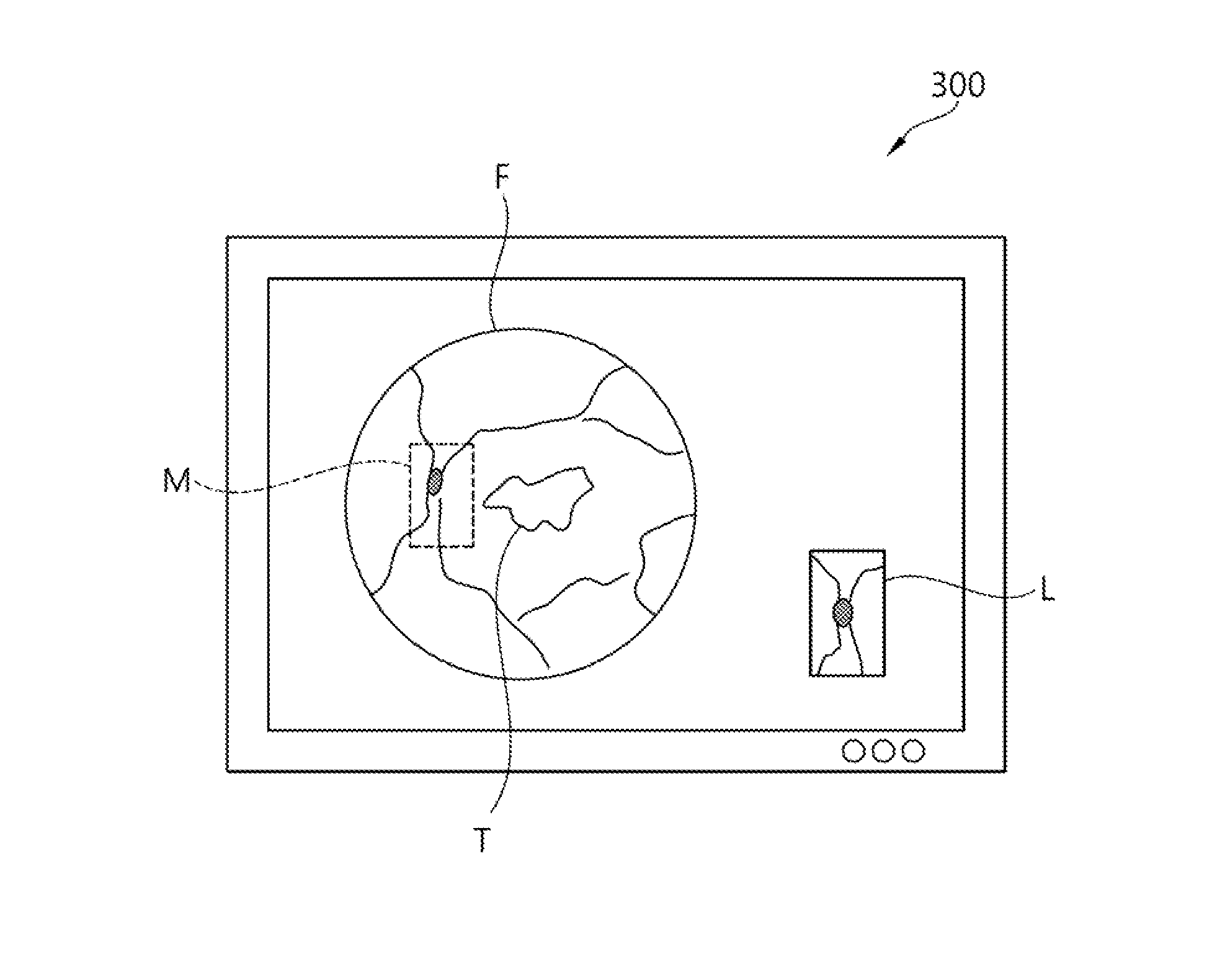
[0049]The method of measuring, by the ophthalmic apparatus 10 configured as described above, a treatment location in accordance with the present invention is described below with reference to FIG. 3.
[0050]First, the first image unit 100 operates and generates the image F of a fundus oculi region of the retina by photographing the fundus oculi region of the retina. The generated image F of the fundus oculi region of the retina is analyzed (S10). Information about a specific region of the image F of the fundus oculi region of the retina analyzed by the image analysis unit 400 at step ‘S10’ is stored in the memory unit 500 (S30).
[0051]The second image unit 200 generates the image L of a local region of the retina by photographing the local region of the retina that is viewed by an operator (S50). The image L of the local region of the retina is mapped to the mapping location M in the image F of the fundus oculi region of the retina using the information stored in the memory unit 500.
[0...
second embodiment
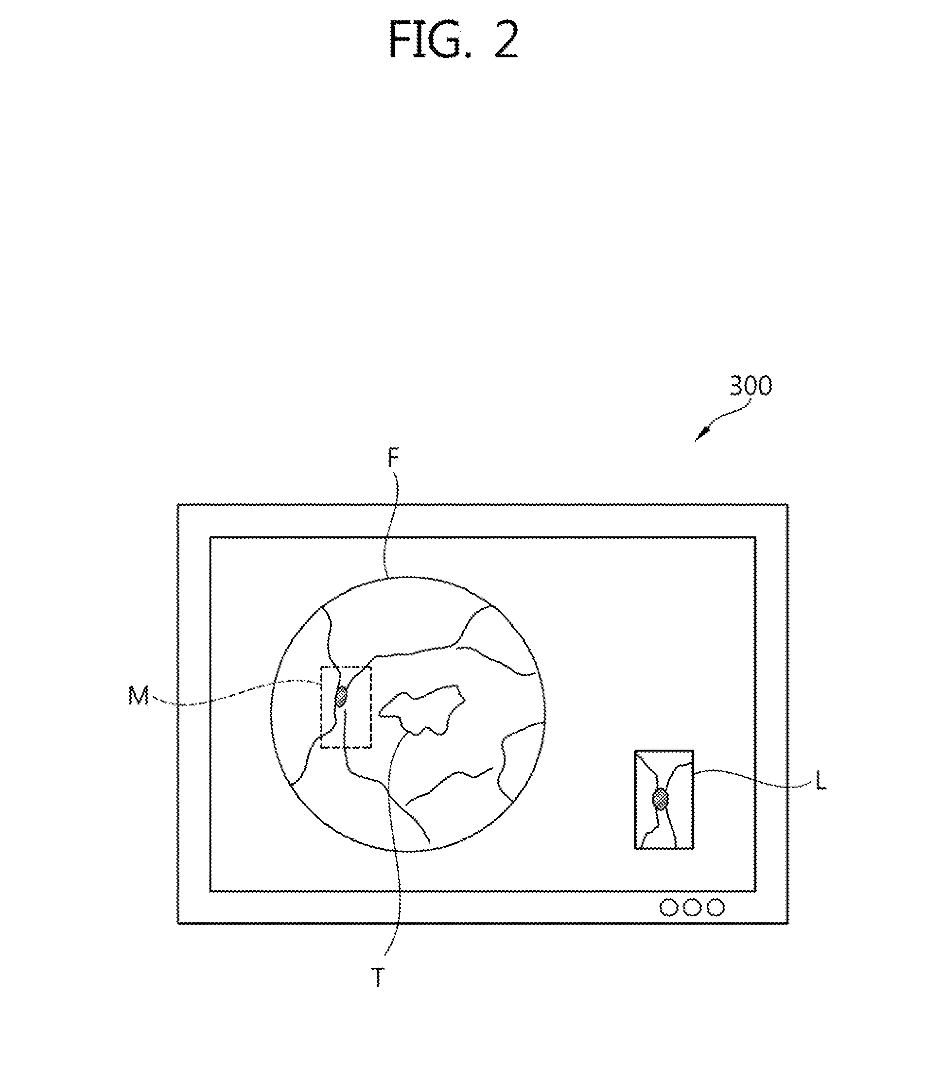
[0056]The method of measuring, by the ophthalmic apparatus 10 configured as described above, a treatment location in accordance with the present invention is described below with reference to FIG. 5.
[0057]First, the first image unit 100 operates and generates the image F of the fundus oculi region of the retina by photographing the fundus oculi region of the retina. The generated image F of the fundus oculi region of the retina is analyzed (S100). Information about a specific region of the image F of the fundus oculi region of the retina analyzed by the image analysis unit 400 at step ‘S100’ is stored in the memory unit 500 (S300).
[0058]The second image unit 200 generates the image L of a local region of the retina by photographing the local region of the retina viewed by an operator (S500). The grids G are displayed in the image F of the fundus oculi region of the retina in order to improve efficiency of the mapping of the image L of the local region of the retina to the mapping lo...
PUM
 Abstract
Description
Claims
Application Information
 - R&D Engineer
- R&D Manager
- IP Professional
- Industry Leading Data Capabilities
- Powerful AI technology
- Patent DNA Extraction
Browse by: Latest US Patents, China's latest patents, Technical Efficacy Thesaurus, Application Domain, Technology Topic.
© 2024 PatSnap. All rights reserved.Legal|Privacy policy|Modern Slavery Act Transparency Statement|Sitemap