
Intermedullary devices for generating and applying compression within a body

a technology of compression device and internal medicine, applied in the field of internal medicine, can solve the problems of slow healing process, device failure to achieve the goal, and disruption of healing tissue,

Active Publication Date: 2015-03-12
ARTHREX
View PDF33 Cites 70 Cited by
  • Summary
  • Abstract
  • Description
  • Claims
  • Application Information

AI Technical Summary

Benefits of technology

The present invention provides better ways to attach bone fragments together, create compression, and keep it for a long time while healing occurs. Specifically, this involves the use of new devices that can grip bones securely and apply a compressive force. Additionally, a second bone-engaging feature is formed on the shaft of the device, which can prevent the shaft from being easily removed from the bone fragment. Overall, this creates a stronger and more stable attachment of bones.

Problems solved by technology

Furthermore, gaps between bone fragments that are too large allow motion to occur between the bone fragments, disrupting the healing tissue and thus slowing the healing process.
While these intramedullary devices are designed to bring the bone fragments into close contact and to generate a compressive load between the bone fragments, these devices do not always succeed in accomplishing this objective.
Furthermore, gripping the bone with only a pair of co-planar legs does not provide significant torsional stability to the fusion site.

Method used


Image

  • Intermedullary devices for generating and applying compression within a body
  • Intermedullary devices for generating and applying compression within a body
  • Intermedullary devices for generating and applying compression within a body

Examples


Embodiment Construction

[0053]The present invention comprises the provision and use of novel intramedullary devices that are better able to bring bone fragments into close proximity with each other, generate a compressive load, and maintain that compressive load for a prolonged period of time while healing occurs.

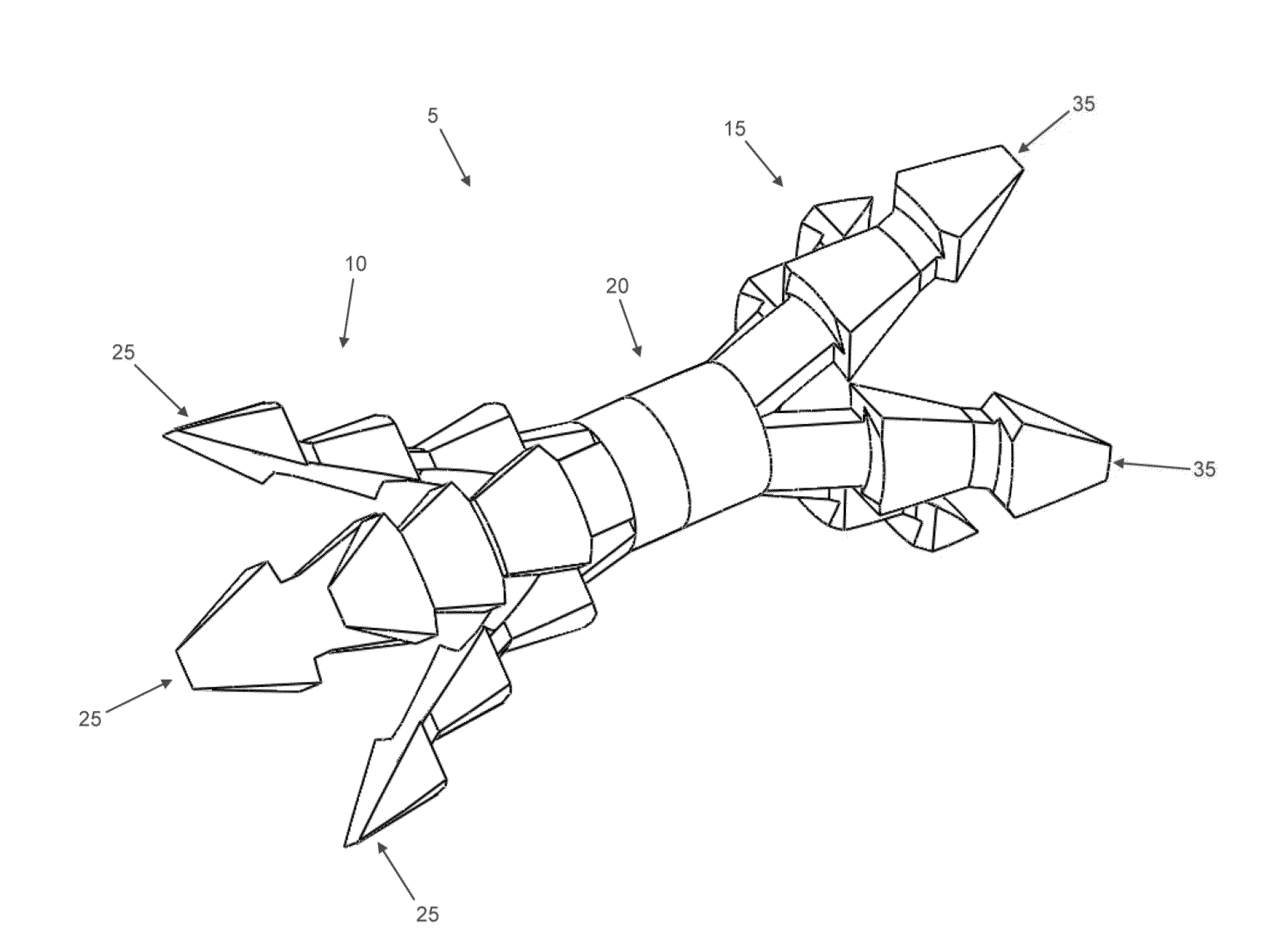
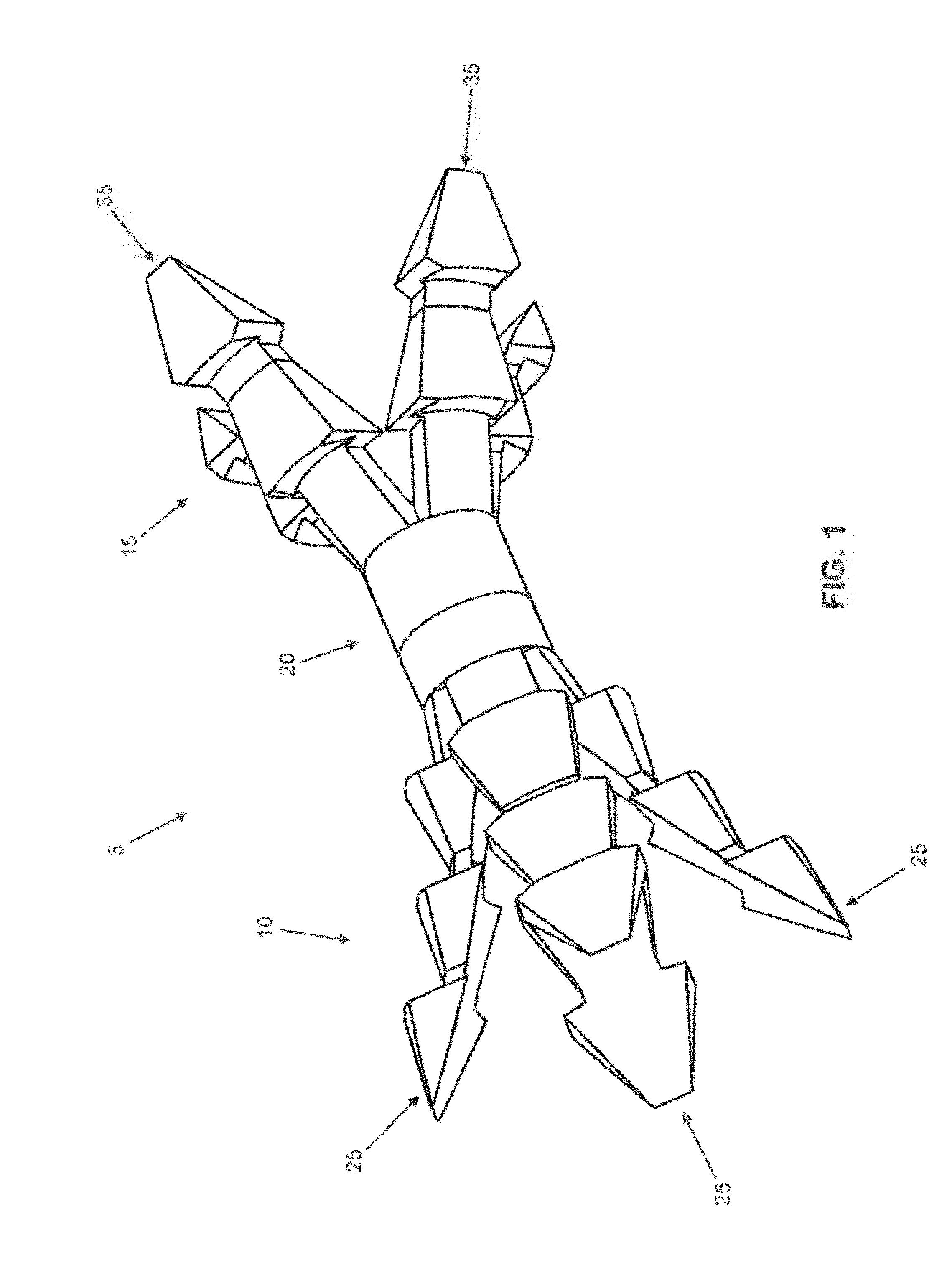
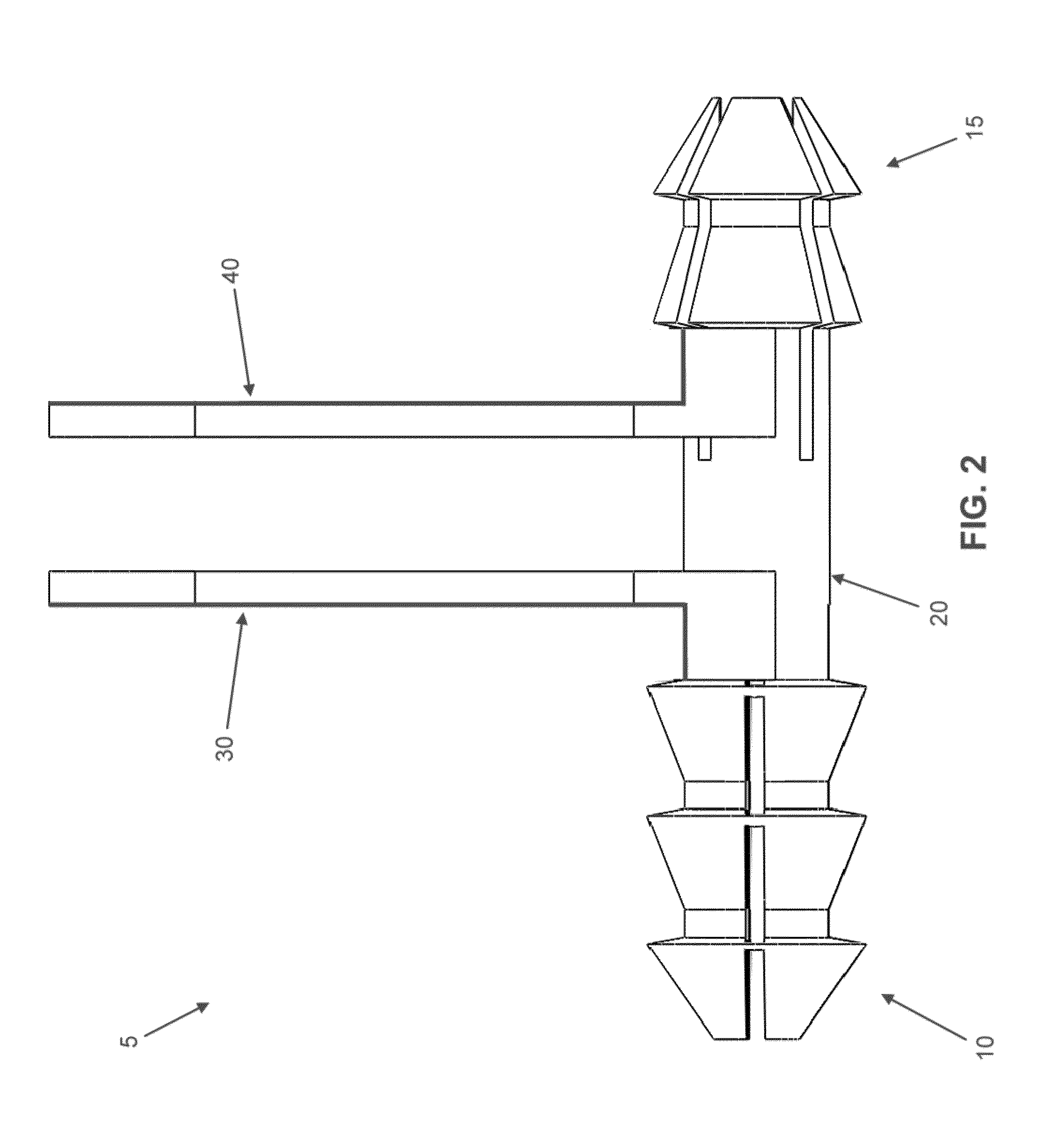
[0054]Looking first at FIGS. 1-4, there is shown an intermedullary fusion device 5 manufactured from a shape memory material (e.g., a material capable of exhibiting superelasticity and / or a temperature-induced shape change). The shape memory material may be a metal alloy (e.g., Nitinol) or a polymer (e.g., appropriately processed PEEK). Intramedullary fusion device 5 comprises a first barbed end region 10, a second barbed end region 15, and a central bridge region 20 connecting first barbed end region 10 to second barbed end region 15. Intramedullary fusion device 5 is preferably cannulated so as to allow the intramedullary fusion device to be installed over a k-wire if desired, while also allowin...


PUM


Abstract

Apparatus for securing a first bone fragment to a second bone fragment, said apparatus comprising: a fusion device, said fusion device comprising: a shaft having a first end and a second end; a first bone-engaging feature formed on said shaft at a first location, said first bone-engaging feature comprising at least one barb which, in its unbiased condition, flares outwardly from the longitudinal axis of said shaft and which is capable of being elastically constrained to a position substantially parallel to the longitudinal axis of said shaft, such that said first end of said shaft may be advanced into a hole in the first bone fragment when said at least one barb is elastically constrained to a position substantially parallel to the longitudinal axis of said shaft but is prevented from being withdrawn from the hole in the first bone fragment when said at least one barb is in its unbiased condition; and a second bone-engaging feature formed on said shaft at a second location, said second bone-engaging feature comprising at least one barb which, in its unbiased condition, flares outwardly from the longitudinal axis of said shaft and which is capable of being elastically constrained to a position substantially parallel to the longitudinal axis of said shaft, such that said second end of said shaft may be advanced into a hole in the second bone fragment when said at least one barb is elastically constrained to a position substantially parallel to the longitudinal axis of said shaft, but is prevented from being withdrawn from the hole in the second bone fragment when said at least one barb is in its unbiased condition.

Description

REFERENCE TO PENDING PRIOR PATENT APPLICATIONS[0001]This patent application:[0002](1) is a continuation-in-part of pending prior U.S. patent application Ser. No. 13 / 624,643, filed Sep. 21, 2012 by MX Orthopedics, Corp. and Matthew Fonte for OSTEOSYNTHETIC SHAPE MEMORY MATERIAL INTRAMEDULLARY BONE STENT AND METHOD FOR TREATING A BONE FRACTURE USING THE SAME (Attorney's Docket No. FONTE-091014), which patent application:[0003](a) claims benefit of prior U.S. Provisional Patent Application Ser. No. 61 / 537,766, filed Sep. 22, 2011 by Matthew Fonte for OSTEOSYNTHETIC SHAPE MEMORY MATERIAL INTRAMEDULLARY BONE STENTS AND METHODS FOR TREATING BONE FRACTURES USING THE SAME (Attorney's Docket No. FONTE-10 PROV); and[0004](b) claims benefit of prior U.S. Provisional Patent Application Ser. No. 61 / 570,091, filed Dec. 13, 2011 by Matthew Fonte for OSTEOSYNTHETIC SHAPE MEMORY MATERIAL INTRAMEDULLARY BONE STENTS AND METHODS FOR TREATING BONE FRACTURES USING THE SAME (Attorney's Docket No. FONTE-14...

Claims


Application Information

Patent Timeline
IPC IPC(8): A61B17/72
CPCA61B17/7266A61B2017/00946A61B2017/00867A61B17/7291
Inventor PALMER, MATTHEWFONTE, MATTHEWDEVANEY, ROBERT
Owner ARTHREX
Learn More