
Laparoscopic surgery

a laparoscopic surgery and laser technology, applied in the field of laparoscopic surgery, can solve the problems of reducing the advantages of laparoscopic surgery, reducing the effect of laparoscopic surgery, and reducing the benefit of laparoscopic surgery

Active Publication Date: 2013-09-26
COLLINS JUSTIN
View PDF2 Cites 84 Cited by
  • Summary
  • Abstract
  • Description
  • Claims
  • Application Information

AI Technical Summary

Benefits of technology

This patent describes a method for safely removing large specimens from the body during laparoscopic surgery. The method involves mechanically morcellating the specimen under irrigation in a confined space. The use of irrigation is advantageous as it maintains the bag in a fully open position and prevents the morcellator from accidentally coming into contact with the bag's sides. The constant circulation of liquid also allows the specimen to rotate away from the bag, ensuring safe and efficient morcellation. In addition, the patent suggests using a laparoscopic bag with multiple layers, one of which is resistant to morcellation to prevent any escape of the specimen. The bag also includes a clear layer to improve visibility for the surgeon and reflect light. The layers have different colorings to alert the surgeon to any breech in the middle layer.

Problems solved by technology

However, one of the limitations of MIS concerns the removal of a relatively large specimen.
Hence, this reduces the advantages of using laparoscopic surgery, which are listed above, and in some cases means that there is actually minimal benefit in performing laparoscopic surgery, over traditional open surgery.
A major concern with this approach is that it is not always possible to ensure that every trace of the morcellated specimen is removed.
Where the specimen is benign, leaving a part of the specimen in the body cavity may lead to infection as the tissue breaks down and acts as a source for infection.
There is even more risk involved in this method when the specimen is malignant, since any escape of malignant cells can lead to tumour seeding.
In addition, since the surgical plume travels throughout the surgical site, metastasis can occur at any point where ‘raw’ areas are located, such as any of the port sites.
While the use of Endo-Bags can reduce the risks, Endo-Bags do not enclose the whole surgical plume, particularly when they are closed only by means of a drawstring at one end.
Therefore, the risk of tumour seeding and port site metastasis remains.
This possibility also presents the danger of tumour seeding.

Method used


Image

  • Laparoscopic surgery
  • Laparoscopic surgery
  • Laparoscopic surgery

Examples


Embodiment Construction

)

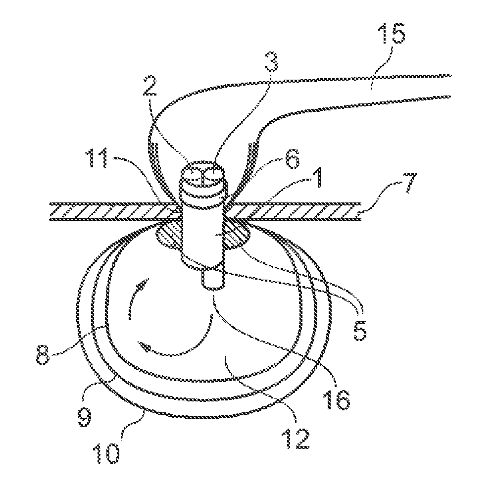
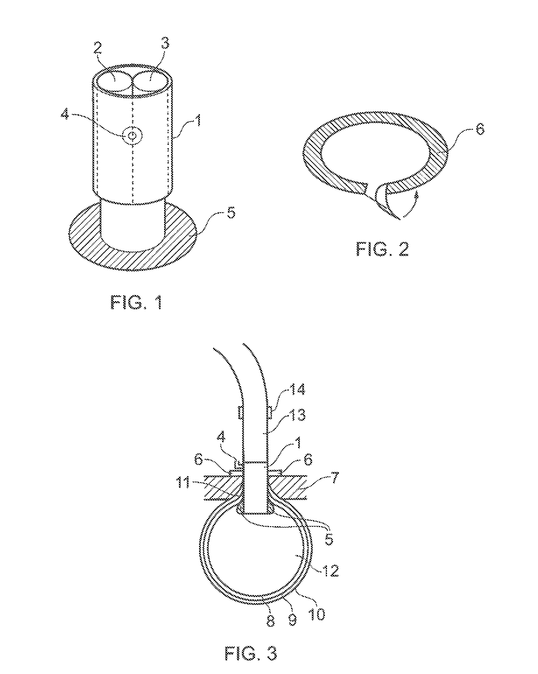
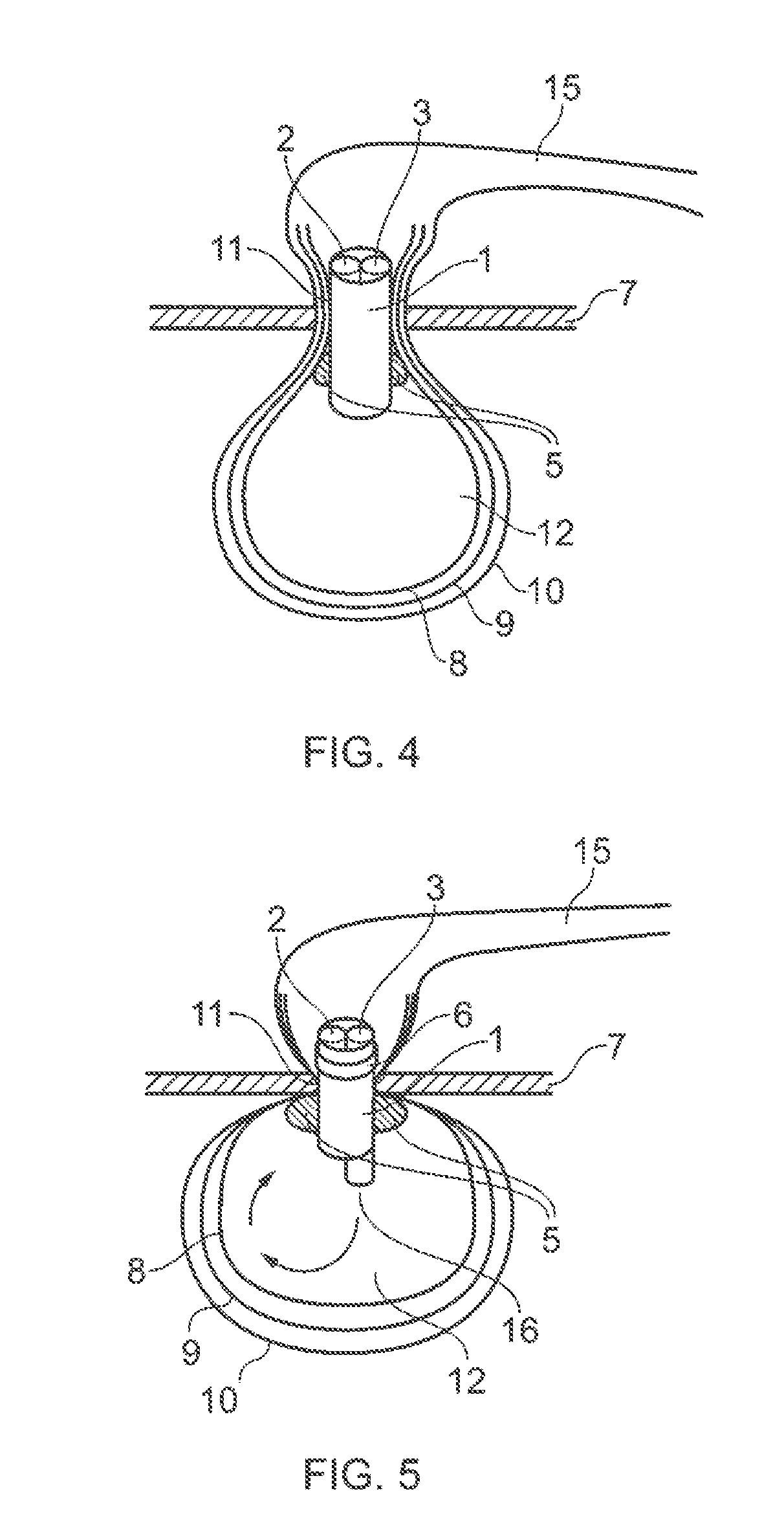
[0072]FIG. 1 shows the tubular housing 1 of a laparoscopic port, through which there are two channels, 2 and 3. The housing 1 is oval in cross section, with a largest diameter of around 20 cm. The housing has an insufflation nozzle, 4, at its proximal end which is in fluid communication with the inflatable cuff, 5. The locking ring 6, shown in FIG. 2, can be passed over the proximal end of the housing 1 and secured next to the skin 7, as shown in FIG. 3. The function of the port is to make a watertight seal around the incision, to hold the bag in place and allow to access for the morcellator, tools for morcellation such as the nephroscope and irrigation tubes.

[0073]FIGS. 3, 4 and 5, show the laparoscopic bag of a preferred embodiment of the present invention with inner layer 8, middle layer 9, and outer layer 10. A bag having 2, or 4 or more layers can also be used in the method of the invention.

[0074]In FIGS. 3, 4 and 5, the laparoscopic port housing 1 is through the skin 7 of a p...


PUM


Abstract

The present invention relates to a method of laparoscopic surgery, and a kit which could be used in laparoscopic and robotic surgery.

Description

[0001]This application claims the benefit of the filing date of U.S. Appl. No. 61 / 531,968, filed Sep. 7, 2011, the entirety of which is fully incorporated by reference herein.FIELD OF THE INVENTION[0002]The present invention relates to a method of laparoscopic surgery, and a kit which could be used in laparoscopic and robotic surgery.BACKGROUND OF THE INVENTION[0003]Laparoscopic surgery, also known as minimal access, minimally invasive or keyhole surgery, is a modern surgical technique. During laparoscopic surgery operations are performed through small incisions, usually 0.5 to 1.5 cm in length. This is in contrast to traditional open surgery where a much longer incision would be made to perform the same operation.[0004]Minimal access surgery also known as minimally invasive surgery (MIS) which includes laparoscopic and robotic surgery and which utilises minimal sized incisions is becoming increasingly popular, with more and more routine operations such as nephrectomy and cystectomy...

Claims


Application Information

Patent Timeline
Patent Type & Authority Applications(United States)
IPC IPC(8): A61B17/221A61B1/307A61B1/018A61B46/23
CPCA61B17/221A61B1/018A61B1/307A61B1/3132A61B2017/00287A61B2017/3445A61B2017/3486A61B17/3421
Inventor COLLINS, JUSTIN
Owner COLLINS JUSTIN
Learn More