
Graphical user interface for operating an MRI

Inactive Publication Date: 2013-03-28
ASPECT IMAGING
View PDF8 Cites 57 Cited by
  • Summary
  • Abstract
  • Description
  • Claims
  • Application Information

AI Technical Summary

Benefits of technology

The invention is a method for scanning using an MRI and a computerized operating system. The method involves displaying a GUI on a display and interacting with the MRI to scan an object. The GUI allows for the selection of different types of objects, their orientation, and the preferred scanning region and quality. The system can also select the type of object and display it in different ways, such as a single image display, multi-image display, thumbnail display, or comparative display. The invention also includes a save scan option and an export option for saving or comparing different scanning sessions. The technical effects of the invention include improved accuracy and efficiency in scanning and the ability to scan a variety of objects and their characteristics.

Problems solved by technology

In addition, handling the images produced by the MRI is done in interfaces which are not intuitive and are limited in their options.
U.S. Pat. No. 5,606,258 describes a controlling system for operating MRI, however, this controlling system still requires the user knowledge of the MRI structure.
However, the user interface is adapted to provide view of single scans and is nor adapted for more complicated requirement such as scans compare.

Method used


Image

  • Graphical user interface for operating an MRI
  • Graphical user interface for operating an MRI
  • Graphical user interface for operating an MRI

Examples


example 1

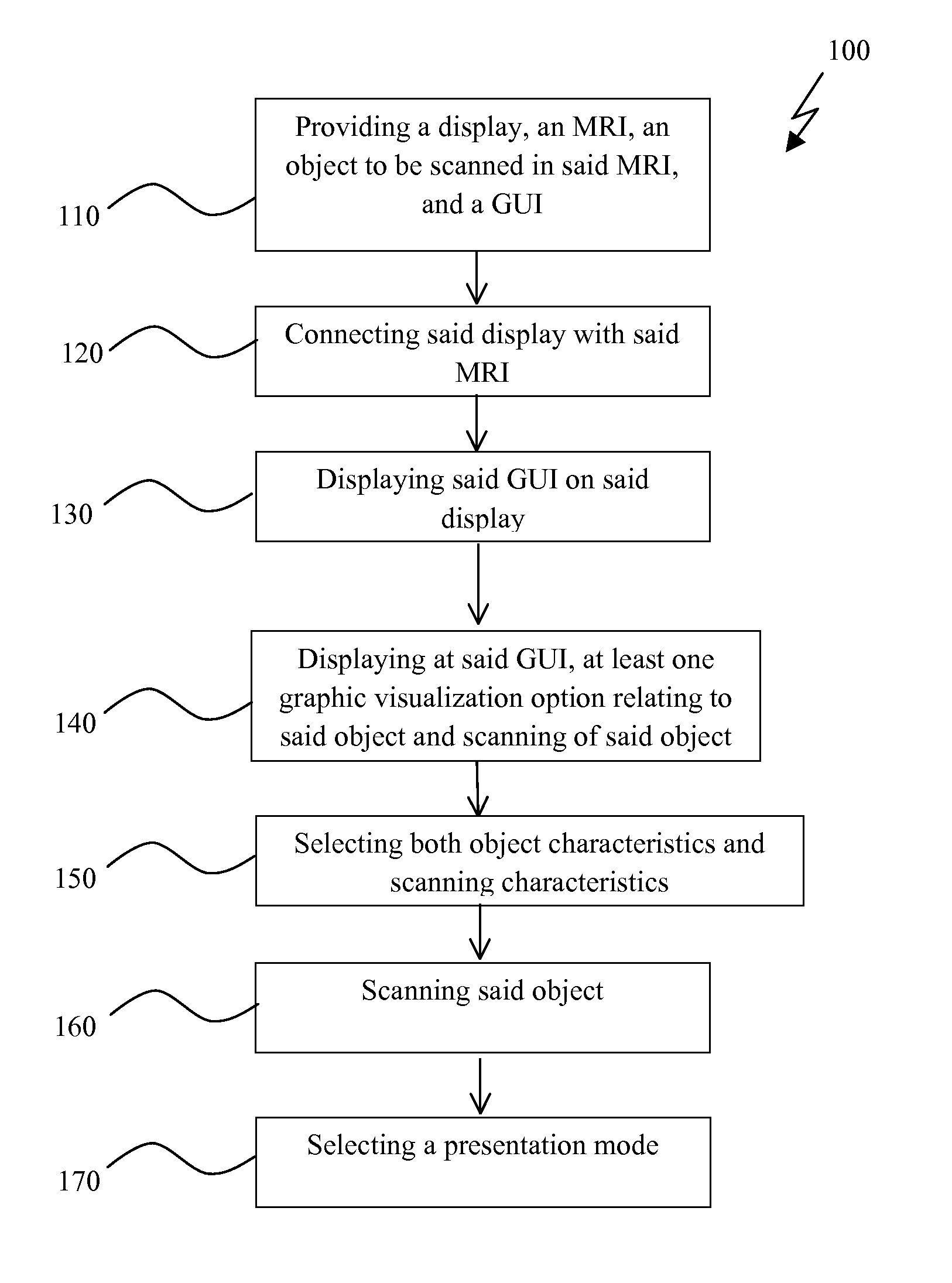
[0077]This example is provided in a non-limiting manner to illustrate one scope of the invention, wherein method 100 includes, inter alis, steps as follows (i) providing a 24 inch size screen equipped with the commercially available operating system named windows XP, with screen resolution of 1920×1200, the commercially available compact MRI system named M2 Compact MRI system, a rat and a GUI 104; (ii) connecting the screen to the M2 and displaying GUI 104 on the display; (iii) presenting on GUI 104 options for selecting type of experiment, a mouse or rat option and the identification number of the rat; (iv) presenting on GUI 104 additional option for selecting whether the rat is in prone position or in supine position; (v) presenting on GUI 104 additional option for selecting tumor location of the rat to be for example either one of head, body or whole body; (vi) presenting on GUI 104 an additional option for selecting image quality for example according to large tumor or small tum...

example 2

[0079]Another example is presented in a non-limiting manner to illustrate the scope of the invention, wherein method 100 includes an MRI adapted to scan humans. Scans are used not only for research purposes but also for diagnostic purposes. In the current example, the method 100 additionally comprising a step of selecting human gender, age and medical history. Additionally, the method comprises a set of steps useful for acquiring treatment history and suggesting future treatment in accordance with scanning results.

[0080]The MRI computerized operating system is now discussed. Reference is made again for FIG. 12, which presents in a non-limiting manner an MRI 102 computerized operating system for scanning object 105. System comprises (i) an MRI 102; (ii) a display 103; (iii) a GUI 104; and, (iv) object 105 to be scanned by MRI 102. Display 103 is operatively connected to MRI, and GUI 104 is presented on display 103. Additionally, GUI 104 presents graphic visualization options relating...

example 3

[0097]This example is presented in a non-limiting manner to illustrate the scope of the invention, wherein in one embodiment of the current invention the system comprises (i) a 24 inch size screen equipped with the commercially available operating system named windows XP, with screen resolution of 1920×1200; (ii) the commercially available compact MRI system named M2 Compact MRI system; (iii) a rat; and (iv) a GUI 104. It is possible to connect the screen to the M2 and to display a GUI on the display. Additionally, GUI 104 presents options for selecting type of experiment, the object identity to be a mouse or rat, and the identification number of the mouse or rat. In this system GUI 104 additional comprises an option for selecting whether the rat is in prone position or in supline position. It is also possible to select via GUI 104 tumor location of the rat to be either one of head, body or whole body. Additionally, GUI 104 comprises an option for selecting image quality in accordan...


PUM


Abstract

A method for scanning using an MRI, including: providing a display, MRI, object to be scanned in said MRI, and a GUI; connecting said display with said MRI; displaying said GUI on said display; displaying at at least one first region of said GUI, graphic visualization options relating to said object and scanning of object, said visualizations are selected from a group consisting of type of object, object orientation, preferred scanning region of object, preferred scanning quality of object, preferred scanning orientation, preferred scanning type, and a combination thereof; selecting said object characteristics and scanning characteristics via said GUI; scanning said object by means of said MRI in accordance with said determined characteristics; and optionally, selecting a presentation mode of said scanning from a group consisting of single image display, multi-image display, thumbnail display, comparative display and a combination thereof, and displaying the same on said display.

Description

FIELD OF THE INVENTION[0001]The present invention generally pertains to the field of MRI operation. In particular, it relates to means and method for operating a user-friendly GUI adapted for controlling MRI parameters and viewing MRI scans.BACKGROUND[0002]Operation of MRI is performed today by directly controlling the MRI parameters; therefore, an MRI user must be a person with extensive knowledge of the physics of MRI. Often, MRI users are researchers which have not been trained to operate an MRI and are in need to be able to control the MRI scanning by a simple non-technical language. In addition, handling the images produced by the MRI is done in interfaces which are not intuitive and are limited in their options.[0003]U.S. Pat. No. 5,606,258 describes a controlling system for operating MRI, however, this controlling system still requires the user knowledge of the MRI structure. US patent application 20100037182 describes a user interface for displaying MRI images. However, the ...

Claims


Application Information

Patent Timeline
IPC IPC(8): A61B5/055
CPCG01R33/546
Inventor RAPOPORT, URI
Owner ASPECT IMAGING
Learn More