
Device and method for assisting in initial setting of imaging condition, and radiation imaging apparatus

a radiation imaging apparatus and imaging condition technology, applied in the direction of material analysis using wave/particle radiation, instruments, applications, etc., can solve the problems of difficult simulation of imaging conditions, method disclosed, and ineffectiveness of u.s. patent no. 7,949,098, and achieve the effect of reducing the dos

Inactive Publication Date: 2013-03-28
FUJIFILM CORP
View PDF19 Cites 11 Cited by
  • Summary
  • Abstract
  • Description
  • Claims
  • Application Information

AI Technical Summary

Benefits of technology

The present invention provides a device and method to help set the initial settings for a radiation imaging apparatus. This device can detect the image quality of the image detection panel and adjust the imaging condition accordingly, to minimize the amount of radiation needed for the image. This improves the performance of the imaging apparatus and reduces the risk of radiation exposure to patients.

Problems solved by technology

However, the method disclosed in the U.S. Pat. No. 7,949,098 is not effective when an image detection panel of a new model is introduced instead of the model currently used, for example, typically, when a medical institution using the IP cassette introduces an electronic cassette.
It is difficult to simulate the imaging condition for the new image detection panel through image processing of the past image captured with the existing image detection panel.
As a result, effective use of the new image detection panel is hampered, for example, the imaging condition corresponding to the existing image detection panel may be applied to the new image detection panel with high sensitivity even though the new image detection panel can be used in a different imaging condition with a reduced dose.
However, because the preliminary imaging for determining the imaging condition exposes the patient to unnecessary radiation, the preliminary imaging cannot be employed in view of dose reduction.
The U.S. Pat. No. 7,949,098 does not disclose or even suggest these problems and their solutions.

Method used


Image

  • Device and method for assisting in initial setting of imaging condition, and radiation imaging apparatus
  • Device and method for assisting in initial setting of imaging condition, and radiation imaging apparatus
  • Device and method for assisting in initial setting of imaging condition, and radiation imaging apparatus

Examples


first embodiment

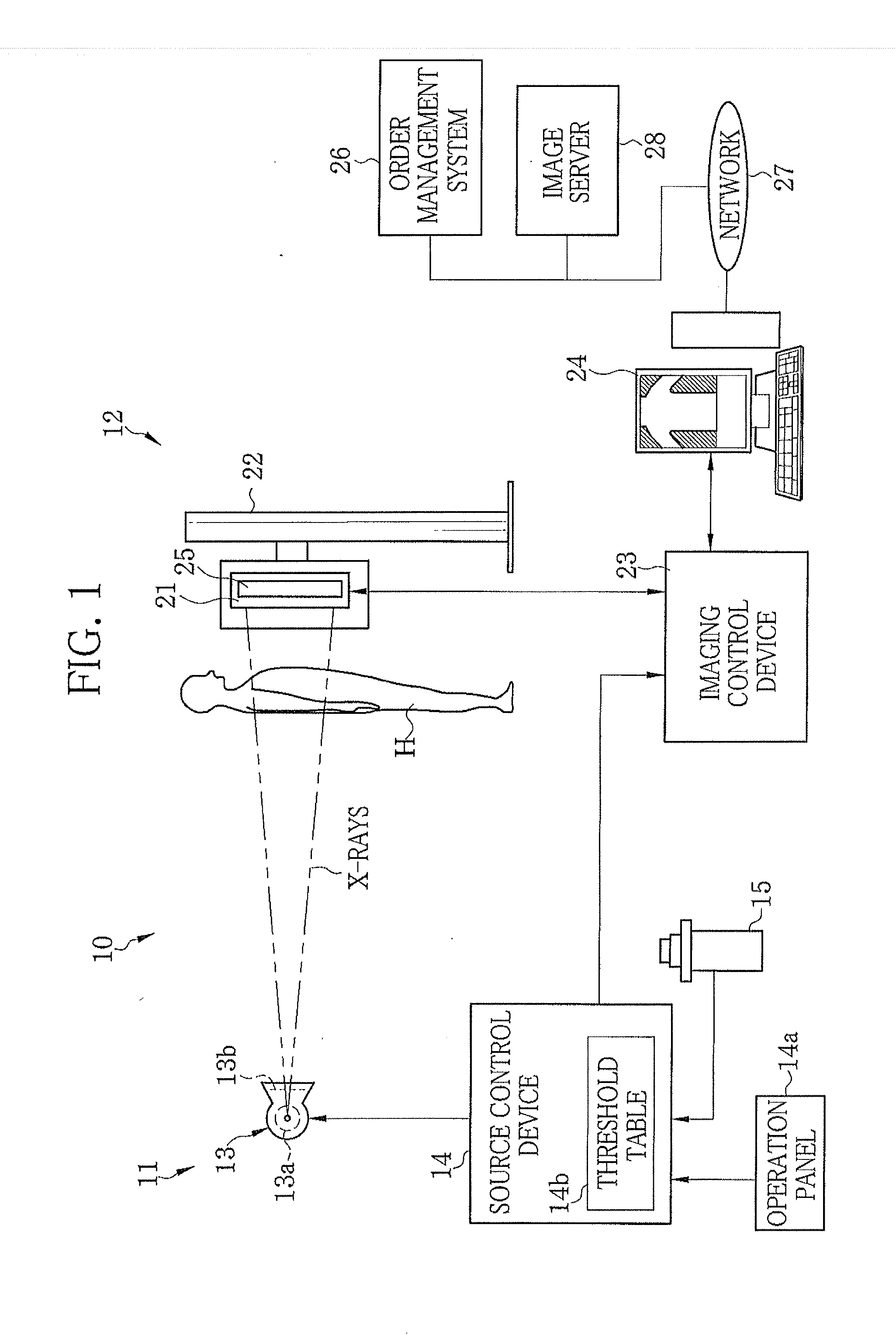
[0049]In FIG. 1, a radiation system, for example, an X-ray imaging system 10 is composed of an X-ray generating device 11 and an X-ray imaging apparatus 12. The X-ray generating device 11 is composed of an X-ray source 13, a source control device 14 for controlling the X-ray source 13, and a radiation switch 15. The X-ray source 13 has an X-ray tube 13a for emitting X-rays and a collimator 13b for limiting an X-ray field of the X-rays emitted from the X-ray tube 13a.

[0050]The X-ray tube 13a has a cathode and an anode (target). The cathode is composed of a filament that releases thermoelectrons. The target emits the X-rays when struck by the thermoelectrons released from the filament. The collimator 13b has a plurality of lead plates arranged in a lattice-like pattern to shield the X-rays. An opening for passing the X-rays is formed at the center of the lattice-like lead plates. The size of the opening is varied by moving the lead plates. Thus, the X-ray field is limited.

[0051]The s...

second embodiment

[0134]In the second embodiment, an additional function is provided to the X-ray imaging system 10 of the first embodiment. The additional function is to receive the selection instruction of the dose on a doctor-by-doctor basis and display selection statuses of the doctors. The second embodiment is similar to the first embodiment except for the additional function. The additional function is mainly described in the following.

[0135]As shown in FIG. 18, a dose selection screen 91 of the second embodiment is provided with a doctor ID input box 92 and a selection status button 93. A doctor ID, being identification information of a doctor, is inputted to the ID input box 92. The main controller 41b receives the selection instruction of the dose, through the sample image selected, in association with the doctor ID inputted to the doctor ID input box 92. The selection instruction is stored in the memory 42 or the storage device 43.

[0136]When the selection status button 93 is clicked, the ma...

third embodiment

[0139]In a third embodiment, an additional function is provided to the X-ray imaging system of the first or second embodiment. Using the additional function, a result of comparison between a dose used for past image(s) captured with the existing image detection panel (for example, the IP cassette) in the medical institution and a dose used for an image captured with the electronic cassette 21 newly introduced to the medical institution is displayed. The third embodiment is similar to the first or second embodiment except for the additional function. The additional function is mainly described in the following.

[0140]In FIG. 20, when the electronic cassette 21 is newly introduced, the image server 28 contains past images 100 captured using the IP cassette. The main controller 41b obtains two or more past images 100 from the image server 28 to perform statistical processing of the doses of the past images 100 (for example, calculation to obtain an average dose of the past images 100). ...


PUM

PropertyMeasurementUnit
distanceaaaaaaaaaa
tube voltageaaaaaaaaaa
imagingaaaaaaaaaa

Abstract

Sample data is stored in a console of an electronic cassette (image detection panel). The sample data corresponds to a model of the electronic cassette and includes sample images captured using different doses before a shipment of the electronic cassette. In initial setting processing of an imaging condition including the dose, the sample data is read out and a dose selection screen in which the sample images are arranged is displayed on a display. In the dose selection screen, the sample images are arranged in a tiled manner in an ascending or descending order of their doses (densities). A user compares the sample images to select the sample image with the density suitable for diagnosis. The dose corresponding to the selected sample image is set as an initial value.

Description

BACKGROUND OF THE INVENTION[0001]1. Field of the Invention[0002]The present invention relates to a device and a method for assisting in initial setting of an imaging condition in radiation imaging, and a radiation imaging apparatus having a function to assist in initial setting of an imaging condition.[0003]2. Description Related to the Prior Art[0004]In the medical field, imaging apparatuses using radiation, for example, X-rays are known. The X-ray imaging apparatus includes an image detection panel and a console. The image detection panel receives the X-rays, applied from an X-ray source and passed through a subject, to detect an X-ray image. The image detection panel outputs the X-ray image as digital data. The console sets imaging conditions including a dose. Examples of the image detection panel include an IP cassette using an imaging plate (IP) and an electronic cassette using a flat panel detector (FPD). Having advantages of using a computer for image processing and data inst...

Claims


Application Information

Patent Timeline
Patent Type & Authority Applications(United States)
IPC IPC(8): A61B6/00G01N23/083
CPCA61B6/463A61B6/468A61B6/542H05G1/42A61B6/588H05G1/30A61B6/545
Inventor TSUJI, TETSUYA
Owner FUJIFILM CORP
Learn More