Compact probe for tracer-assisted diagnostic and surgery

a tracer-assisted diagnostic and surgical technology, applied in the field of compact probes for tracer-assisted diagnostic and surgery, can solve the problems of difficult to distinguish tumoral areas from healthy tissues, large background, and relatively long radiation range, so as to eliminate all radioactivity risks, reduce costs, and high affinity

Inactive Publication Date: 2012-09-13
FORIMTECH
View PDF3 Cites 2 Cited by
  • Summary
  • Abstract
  • Description
  • Claims
  • Application Information

AI Technical Summary

Benefits of technology

[0007]Positron-emitting isotope markers with a high affinity for cancerous tissues are known, an example being 18F-labeled 2-fluorodeoxy-D-glucose (FDG). 18F-labeled FDG is a specific marker for a carbohydrate hyper metabolism indicative of malignant tissues or inflammatory tissues. This marker is already used in diagnostic medicine for mapping the spread of a cancer with the aid of complex and expensive positron-detecting equipment (PET (Positron Emission Tomography) camera). After diagnostic examination, the 18F-labeled FDG, which is still present in the tumors, can be used for guiding of surgery or biopsy tools towards the tumor. However, this technique has a serious disadvantage—it can be applied only shortly after PET-examination (due to fast decay of 18F) and can be used only in clinics with special equipment (PET scanners and all related radio-protection facilities). Therefore, replacing FDG or other radioactive markers by fluorescent ones can open much wider applications for this technique, reduce costs and eliminate all risks related to radioactivity.

Problems solved by technology

However, g radiation has the disadvantage of a relatively long range within biological tissues, which creates a considerable background.
It is thus difficult to differentiate the tumoral areas from the healthy tissues.
Moreover, this contamination by the g radiation background makes it difficult, if not impossible, to detect small radio-labelled tumoral objects.

Method used


Image

  • Compact probe for tracer-assisted diagnostic and surgery
  • Compact probe for tracer-assisted diagnostic and surgery

Examples


Embodiment Construction

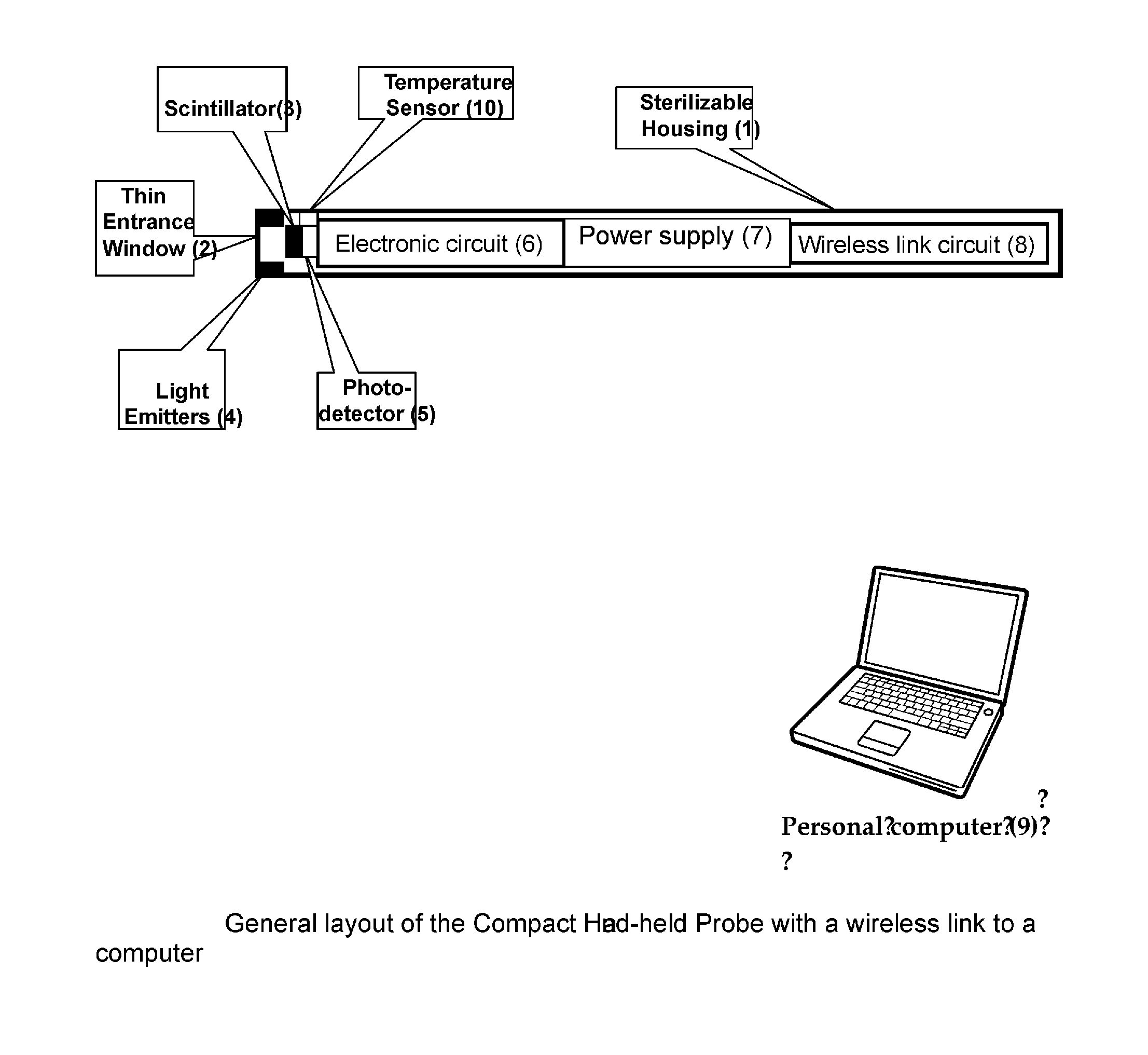
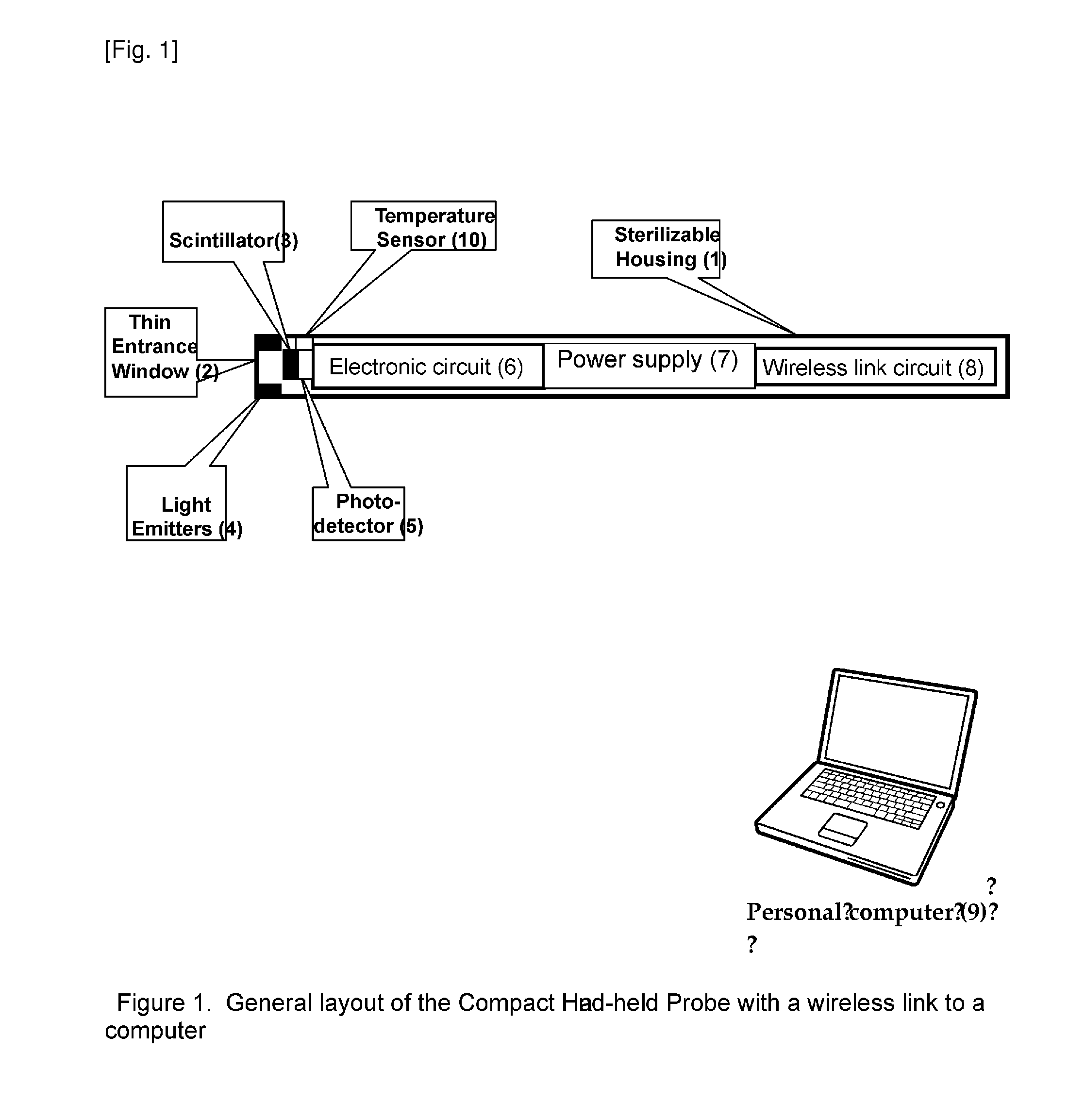
[0009]A new method (claim 1) and device (claims 2 and 3) is proposed which enhances efficiency of procedures using radioactive tracers and allows replace radioactive tracers by more cheap and safe fluorescent ones. The compact probe according to the invention is shown in FIG. 1. Preferred embodiments of the invention are listed in the dependent claims. The probe according to the invention is designed in a way which provides maximum convenience and simplicity in operation for the user, minimal weight and cost (potential disposability), preserving at the same time maximum performance. This is reached by introduction of several novelties.

[0010]The method consists in detection of signals emitted by tracers with time-resolved photon counting technique in a compact, simple and handy autonomous detector, which is supplied in a sterile packaging ready for operation, and transmitting those signals using wireless link to a remote computer (9), which performs most of the functions, such as: co...


PUM


Abstract

A simple and potentially disposable compact probe of FIG. 1, which is primarily aimed to be used for radio-guided or fluorescence-guided surgery, diagnostics or biopsy, and method of using it, is invented. The novel method reduces limitations inherent to the existing technologies. It consists in shifting most of the functions (signal analysis, detector controls and user visual and audio interfaces) from the probe to an external personal computer connected with the probe via wireless link. Maximum simplification and miniaturization of the probe itself makes it potentially disposable, supplied in sterile package similar to disposable syringe. Due to use of single photon time-resolved counting photo-sensors and microelectronic circuits with sub-nanosecond timing, the probe can be used with fluorescent markers as well. New probe should enhance efficiency of medical procedures and open opportunities for more wide application of the intra-operative probing techniques in medical practice, especially in oncology. Applications other than medical are also possible.

Description

STATE OF THE ART AND INVENTION DESCRIPTIONState of the Art[0001]Surgical radio-guided procedures have been used since a long time and consist in injecting patients with a radioactive isotope that emits b radiation (positrons and / or electrons) and / or g radiation and has the property of binding preferentially to the diseased tissues, e.g. the tumors, via their carrier molecules. The surgeon then uses a so-called ‘peroperative’ hand-held probe sensitive to the radioactivity emitted by the radio-isotope carrier molecules.[0002]This type of radio-guided surgery has proven its efficiency and is commonly recognized and employed for the treatment of lung cancers, melanomas, thyroid cancer, neuroendocrine cancer and benign tumors such as, inter alia, parathyroid hyperplasias or osteoid osteomas. On the other hand, this radio-guided surgical technique using radiosensitive peroperative manual probes is still undergoing evaluation for applications in the treatment of tooth neck or colon cancers...

Claims


Application Information

Patent Timeline
Patent Type & Authority Applications(United States)
IPC IPC(8): A61B6/00
CPCA61B6/4258G01T1/161
Inventor GRIGORIEV, EUGENE
Owner FORIMTECH
Why Eureka
Try Eureka