Mesenchymal stem cell differentiation
a stem cell and mesenchymal technology, applied in the field ofmusculoskeletal disorders, can solve the problems of arthritis or joint injury risk, and achieve the effect of reducing the risk of arthritis or joint injury, and improving or preventing arthritis or joint injury
- Summary
- Abstract
- Description
- Claims
- Application Information
AI Technical Summary
Benefits of technology
Problems solved by technology
Method used
Image
Examples
example 1
High Throughput Screen for Inducers of Chondrogenesis
[0075]To identify and define a new, non-invasive strategy for OA joint repair, we developed a non-biased high throughput screen of proteins capable of selectively directing the differentiation of human mesenchymal stem cells (MSCs) to chondrocytes. The assay system models the resident MSCs in cartilage and identifies mediators that stimulate the natural repair potential and enhance integrated cartilage regeneration. We choose to test a unique secreted protein library by a high throughput, cell-based screens of human and mouse MSCs. This approach provides a strategy that allows the rapid identification of uncharacterized and native protein ligands that affect chondrogenesis.
[0076]To study these secreted proteins, two approaches were taken: the production of conditioned media (CM) from a mammalian producer cell line (HEK293T) and the generation of purified proteins from free style HEK-F cells. Together these complementary approaches...
example 2
Expression of Recombinant Full Length ANGPTL3 and Mutant Protein and Function Analysis
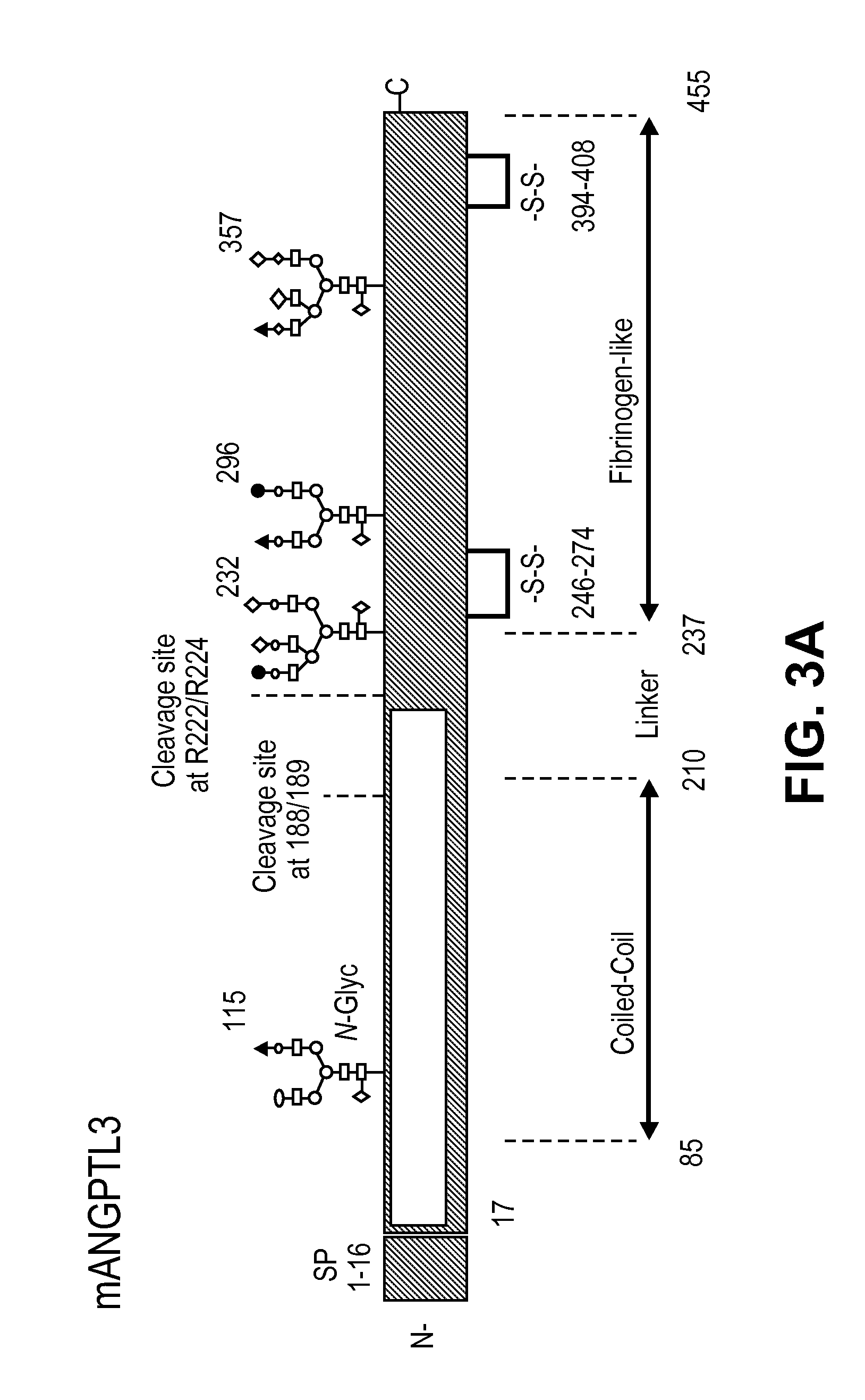
[0081]Mouse ANGPTL3 is predicted to be 51 kDa protein. It belongs to a family of 7 identified Angiopoietin-like (ANGPTL) proteins that have structural similarity to the angiopoiteins, but lack the ability to bind the Tie2 receptor and have distinct functions. They contain an N-terminal coiled-coil domain (CCD) and a C-terminal fibrinogen-like domain (FLD). ANGPTL proteins are tightly regulated by their microenvironment and interactions with the extracellular matrix (ECM), yet the precise interaction sites nor partners have been elucidated in detail. ANGPTL3 is secreted by the liver and circulates systemically. It is controlled through liver X receptors (LXR), with evidence that LXR-induced hyper-triglyceridmedia is due to ANGPTL3 release. Interactions between the CCD and the ECM through a putative heparin binding motif may lead to inhibition of cleavage at the proprotein convertase recognition sequ...
example 3
In vivo Analysis of ANGPTL3
[0086]We have performed several in vivo evaluations of the full length ANGPTL3 to address potential adverse events and intra-articular retention. Following intra-articular (IA) injection of the left knee joints of 8 week old C57BL / 10 mice with 3.6 μg of full length ANGPTL3, we evaluated immunohistochemically the expression in the presence and absence of exposure for 24 hours. By 24 hours, very little to no detectable levels of ANGPTL3 should remain in the synovial fluid as the typical turnover by trans-synovial flow into the synovial lymph vessels for proteins and water is approximately 2 hours. The results revealed no endogenous expression of ANGPTL3 in untreated joints, but significant detection of the protein in the pericellular matrix in the articular cartilage and in the menisci even at the 24 hour time point. Additionally no widespread cytotoxicity to the chondrocytes or cartilage damage in vivo was detected. Following a series of 3 IA injections int...
PUM
| Property | Measurement | Unit |
|---|---|---|
| concentrations | aaaaa | aaaaa |
| mass | aaaaa | aaaaa |
| composition | aaaaa | aaaaa |
Abstract
Description
Claims
Application Information
 - R&D
- Intellectual Property
- Life Sciences
- Materials
- Tech Scout
- Unparalleled Data Quality
- Higher Quality Content
- 60% Fewer Hallucinations
Browse by: Latest US Patents, China's latest patents, Technical Efficacy Thesaurus, Application Domain, Technology Topic, Popular Technical Reports.
© 2025 PatSnap. All rights reserved.Legal|Privacy policy|Modern Slavery Act Transparency Statement|Sitemap|About US| Contact US: help@patsnap.com


