
System Non-invasive Cardiac Output Determination

a cardiac output and non-invasive technology, applied in the field of system for determining cardiac output and stroke volume, can solve the problems of increasing the complexity of clinical procedures, adding to the risk of patients, and different methods, and achieve the effect of reducing the volume of blood flow

Inactive Publication Date: 2012-06-14
SIEMENS HEALTHCARE GMBH
View PDF2 Cites 14 Cited by
  • Summary
  • Abstract
  • Description
  • Claims
  • Application Information

AI Technical Summary

Benefits of technology

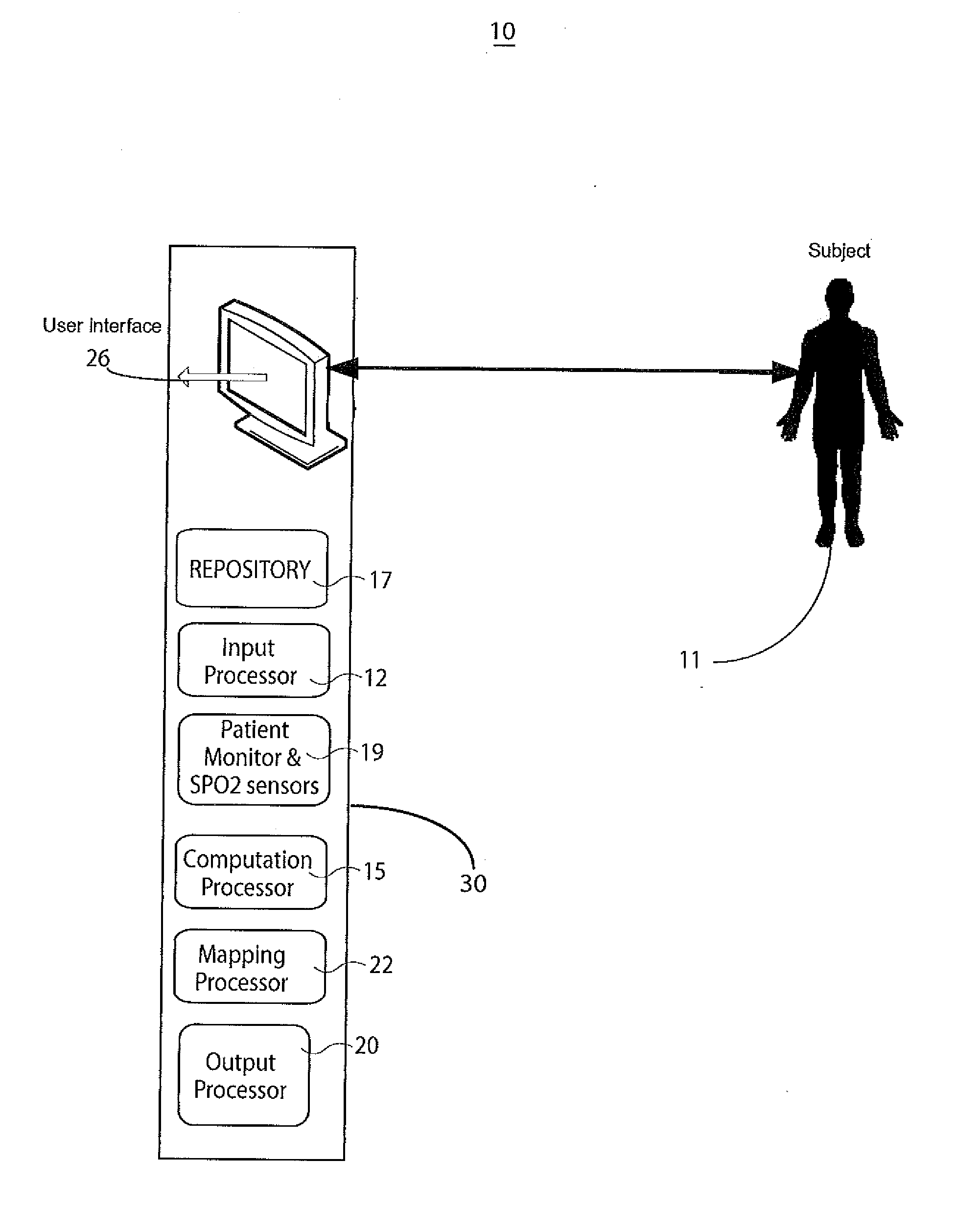
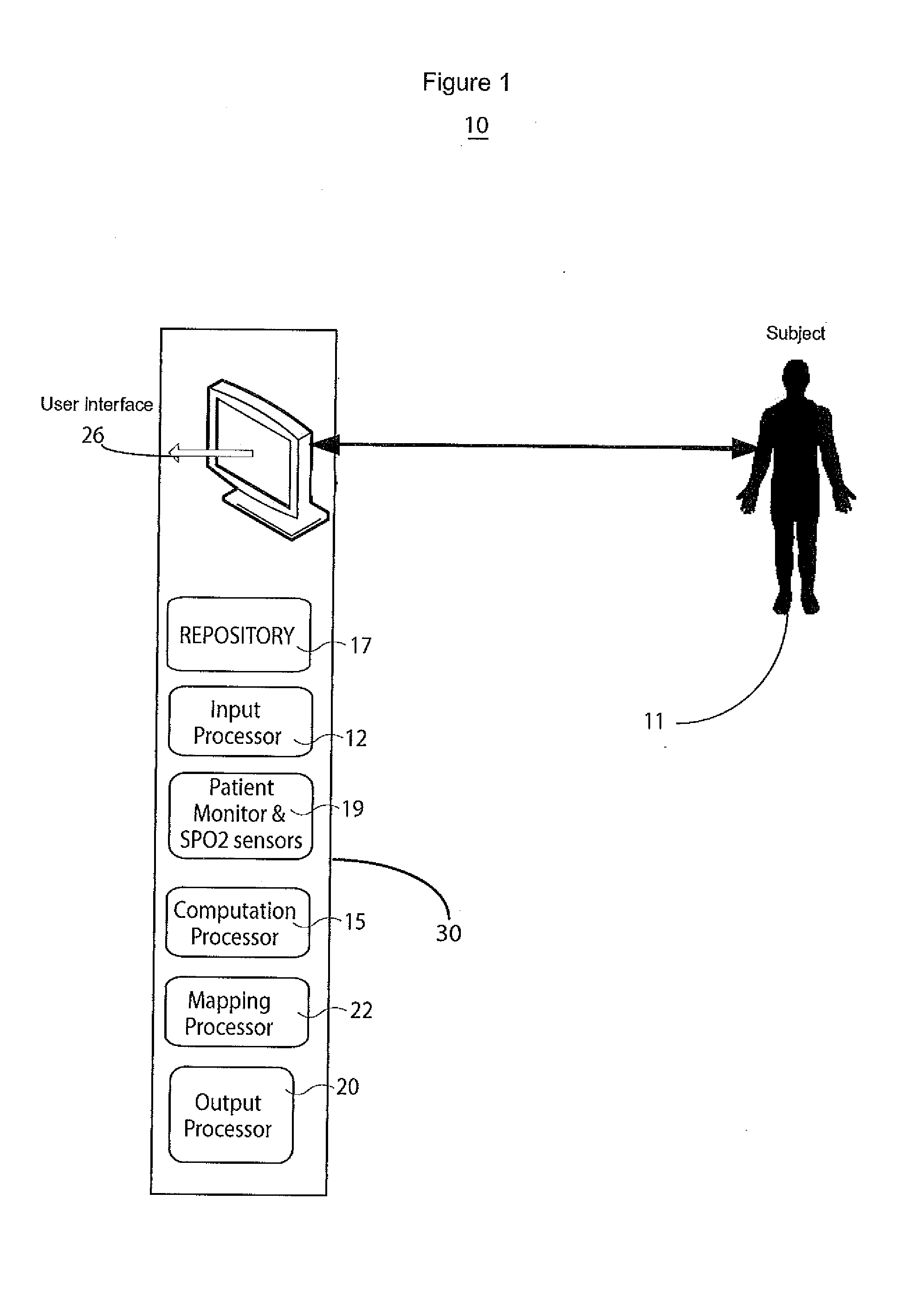
[0005]A system determines cardiac output and stroke volume by using non-invasive oximetric signals, such as SPO2 data and associated waveform, to determine blood flow quantitatively. A non-invasive system determines cardiac output or stroke volume. The system includes an input processor for receiving signal data representing oxygen content of blood of a patient at a particular anatomical location. A computation processor uses the received signal data in calculating a heart stroke volume of the patient comprising volume of blood transferred through the blood vessel in a heart cycle, in response to, a blood volume derived in response to oxygen content of patient blood and at least one factor representing reduction in blood flow volume from a patient heart to the particular anatomical location. An output processor provides data representing the calculated heart stroke volume to a destination device.

Problems solved by technology

However most of these clinical methods are invasive and unreliable which limits their use and results in additional risk to patients.
However these methods have different limitations and disadvantages
Known clinical methods for CO and SV calculation are mostly invasive and require catheters and this adds to clinical procedure complexity and poses additional risk to patients.
The known clinical methods for CO, SV calculation require extensive clinical experience and knowledge for interpretation of the parameters and for calculation accuracy and are also often complex, and time consuming and may be unsuitable for particular clinical environments.
Further known cardiac output calculation methods may be dependent on sensor quality and be sensitive to noise (such as from a power line, patient movement, or treatment, such as pacing and drug delivery) resulting in an unreliable cardiac function calculation.

Method used


Image

  • System Non-invasive Cardiac Output Determination
  • System Non-invasive Cardiac Output Determination
  • System Non-invasive Cardiac Output Determination

Examples


Embodiment Construction

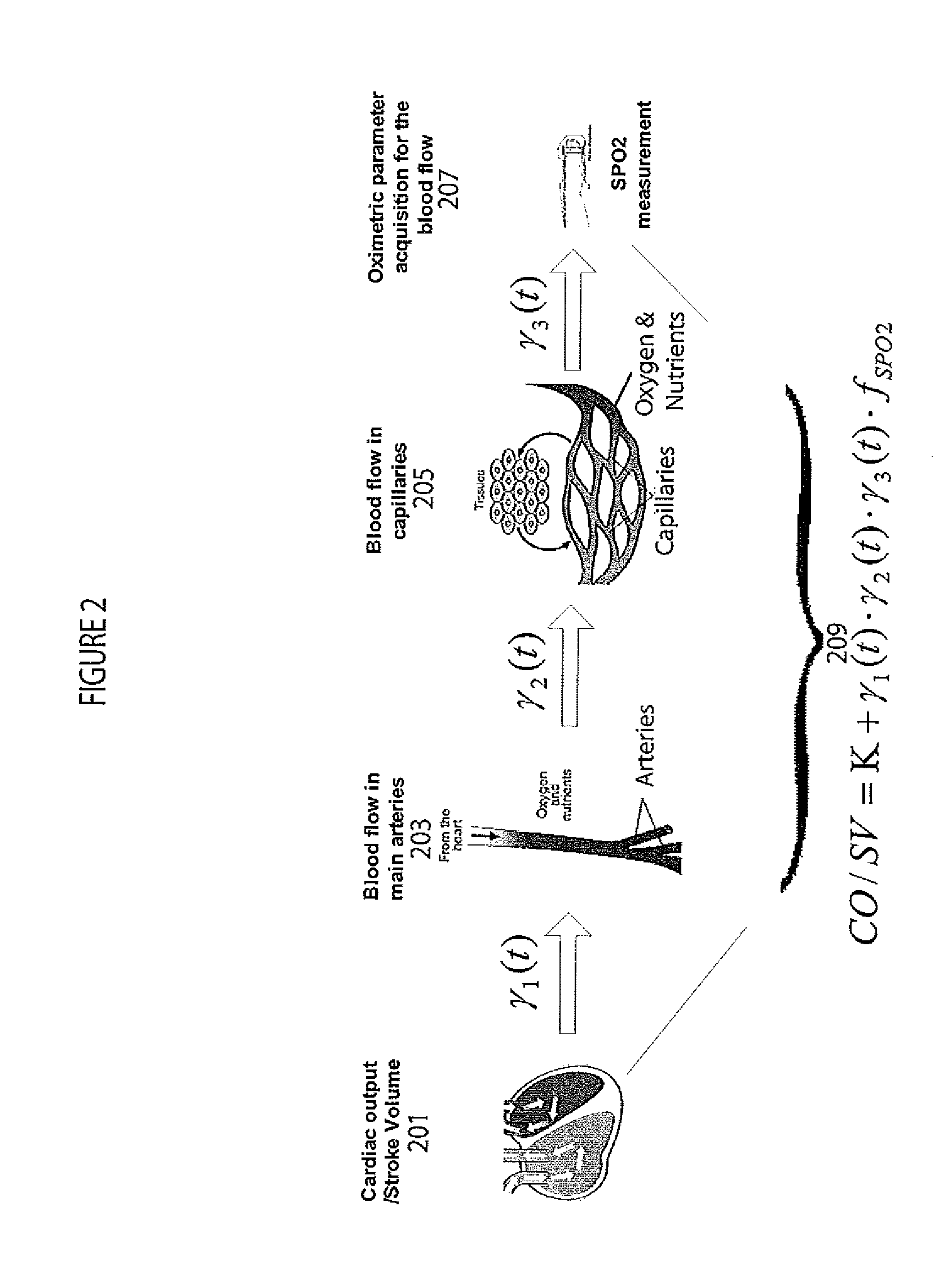
[0013]A system determines cardiac output and stroke volume by using non-invasive oximetric signals, such as blood oxygen saturation (SPO2) data to quantitatively determine blood flow. The SPO2 data is utilized to analyze heart function and blood flow characteristics by building a bridging model between non-invasive blood oximetric signals in capillaries (such as in a finger tip) and cardiac pumps comprising heart chambers (particularly a left ventricle). Using nonlinear modeling based on SPO2 signal properties (such as Density, Variability, Variation), the system accurately determines cardiac output in the presence or absence of substantial noise. The system detects cardiac disorders, differentiates between cardiac arrhythmias, characterizes pathological severity, predicts life-threatening events, and facilitates evaluation of the effects of drug administration to a patient.

[0014]The system quantitatively determines CO and SV values by determining a blood oxygen content (SPO2) repre...


PUM


Abstract

A system determines cardiac output and stroke volume by using non-invasive oximetric signals, such as SPO2 data and waveform, to determine blood flow quantitatively. A non-invasive system determines cardiac output or stroke volume. The system includes an input processor for receiving signal data representing oxygen content of blood of a patient at a particular anatomical location. A computation processor uses the received signal data in calculating a heart stroke volume of the patient comprising volume of blood transferred through the blood vessel in a heart cycle, in response to, a blood volume derived in response to oxygen content of patient blood and at least one factor representing reduction in blood flow volume from a patient heart to the particular anatomical location. An output processor provides data representing the calculated heart stroke volume to a destination device.

Description

[0001]This is a non-provisional application of provisional application Ser. No. 61 / 421,234 filed 9 Dec. 2010, by H. Zhang.FIELD OF THE INVENTION[0002]This invention concerns a system for determining cardiac output and stroke volume in response to, a blood volume derived in response to oxygen content of patient blood and at least one factor representing reduction in blood flow volume from a patient heart to a particular anatomical location.BACKGROUND OF THE INVENTION[0003]Cardiac output (CO) or stroke volume (SV) involve measurements of blood volume ejected by a left ventricle in one minute or in one heart beat and are valuable vital sign signals used for patient health status monitoring. There are multiple methods to calculate CO and SV including using a blood pressure waveform, thermodilution, bio-impedance, a pulse contour or ultrasound, for example. However most of these clinical methods are invasive and unreliable which limits their use and results in additional risk to patients...

Claims


Application Information

Patent Timeline
Patent Type & Authority Applications(United States)
IPC IPC(8): A61B5/0205A61B5/1455
CPCA61B5/0261A61B5/029A61B5/0295A61B5/1455
Inventor ZHANG, HONGXUAN
Owner SIEMENS HEALTHCARE GMBH
Learn More