
Multimodal Ultrasound Training System

Active Publication Date: 2012-03-08
RGT UNIV OF CALIFORNIA
View PDF5 Cites 50 Cited by
  • Summary
  • Abstract
  • Description
  • Claims
  • Application Information

AI Technical Summary

Benefits of technology

[0009]The present inventor has recognized the need to provide an improved VR system and method for providing medical training. In particular, the present inventor has recognized that it would be particularly advantageous to provide a multimodal VR medical training system providing not only VR anatomical images, but also one or more of (a) simulations of images that might be obtained using actual imaging devices (e.g., ultrasound, CT, or MRI imaging systems), and (b) simulations of physical forces or other physical conditions that might be experienced by a physician (or other medical personnel) while performing a procedure.

Problems solved by technology

Today, medical educators are under considerable societal pressure and budgetary constraints to enhance the quality of medical education.
Traditional “learning by doing” models have become less acceptable, particularly where invasive procedures and high-risk care are required.
Given that many medical procedures not only are challenging to perform, but also, if performed improperly, can pose significant risks to patient health and safety, such conventional “see one, do one, teach one” training is not always a preferred method of training.
Complications of this procedure can potentially include misplacement of the line, a collapsed lung or hemorrhage, and statistics show that such complications can occur in between 4 to 15 percent of patients having this procedure.
Thus, while it is desirable that medical professionals performing CVL placements be highly experienced in performing the technique, it is not particularly desirable that medical professionals develop their experience by performing the procedure on actual patients.
However, VR systems are often costly items prohibiting wide scale use in the medical training arena.
Although advantageous in many respects, conventional VR training methodologies are still lacking in certain regards.
To begin with, conventional VR training methodologies have not integrated multiple simulated conventional medical technologies along with textbook style learning.
Conventional VR training methodologies are often cost prohibitive for use by entities with many students or trainees.
High costs have prevented the widespread use of VR technologies for medical education and training.

Method used


Image

  • Multimodal Ultrasound Training System
  • Multimodal Ultrasound Training System
  • Multimodal Ultrasound Training System

Examples


Embodiment Construction

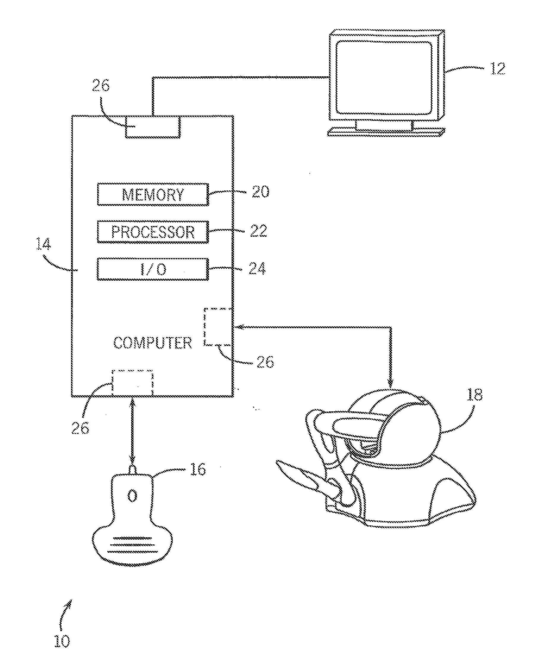
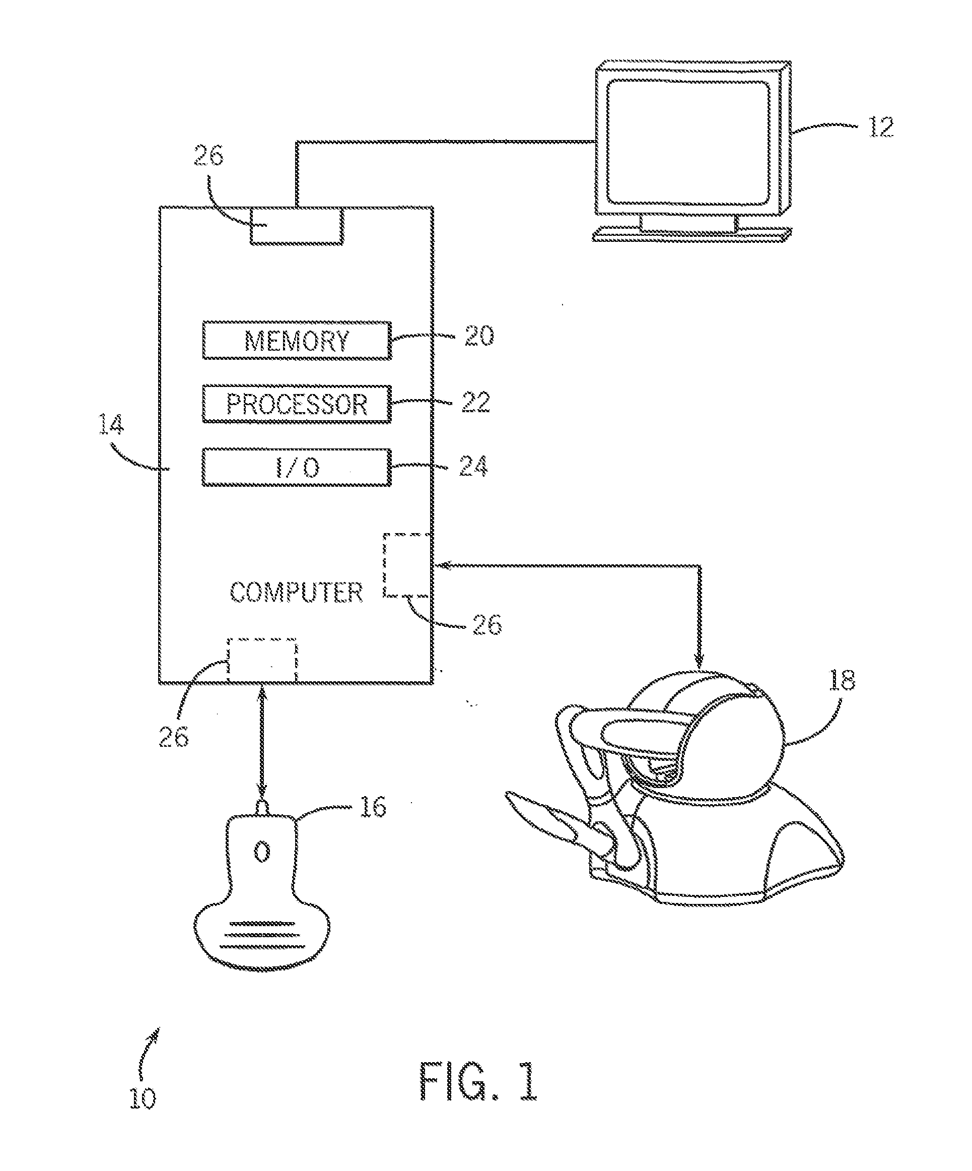
[0020]Referring to FIG. 1, a first exemplary embodiment of an improved medical training system 10 is shown to include a graphical interface 12, a computer 14, a first input / output device 16 and a second input / output device 18. Also as shown, the computer 14 includes a memory device 20, a processor 22, and an input / output device 24. Each of the graphical interface 12, the first input / output device 16, and the second input / output device 18 is connected to the computer 14 by way of conventional connection devices. In the present embodiment, for example, the computer 14 has optional serial ports 26 that serve as interfaces between the computer 12 and each of the graphical interface 12 and the devices 16 and 18. Depending upon the embodiment, the serial ports 26 and other connection component(s) could include any of a variety of different components / devices (e.g., networking components) including, for example, an Ethernet port / link, an RS232 port / communication link, or wireless communica...


PUM


Abstract

The present invention teaches a medical procedure training system based on a PC platform that provides multimodal education within a virtual environment. The system integrates digital video, three-dimensional modeling, and force-feedback devices for the purpose of training medical professionals medical procedures.

Description

CROSS-REFERENCES TO RELATED APPLICATIONS[0001]This patent application is a continuation and claims the benefit of U.S. patent application Ser. No. 11 / 720,515 filed May 30, 2007 for Multimodal Medical Procedure Training System, which claims priority to U.S. Provisional Patent Application No. 60 / 631,488, entitled “Multimodal Emergency Medical Procedural Training Platform” and filed Nov. 30, 2004. Each of those applications is incorporated here by this reference.TECHNICAL FIELD[0002]This invention relates generally to systems and methods for providing medical training, and more specifically to medical training systems and methods that at least partly involve simulations of medical procedures and operations.BACKGROUND ART[0003]Today, medical educators are under considerable societal pressure and budgetary constraints to enhance the quality of medical education. Traditional “learning by doing” models have become less acceptable, particularly where invasive procedures and high-risk care a...

Claims


Application Information

Patent Timeline
IPC IPC(8): G09B23/28
CPCG09B23/286
Inventor SAVITSKY, ERIC A.
Owner RGT UNIV OF CALIFORNIA
Learn More