Pvii phage display
a phage and display technology, applied in the field of phage display, can solve the problems of high probability of destroying the functionality of both piii fusions and the inability of pvii to be used for phage display
- Summary
- Abstract
- Description
- Claims
- Application Information
AI Technical Summary
Problems solved by technology
Method used
Image
Examples
example 1
Modified Helper Phages with Peptides Fused to pVII
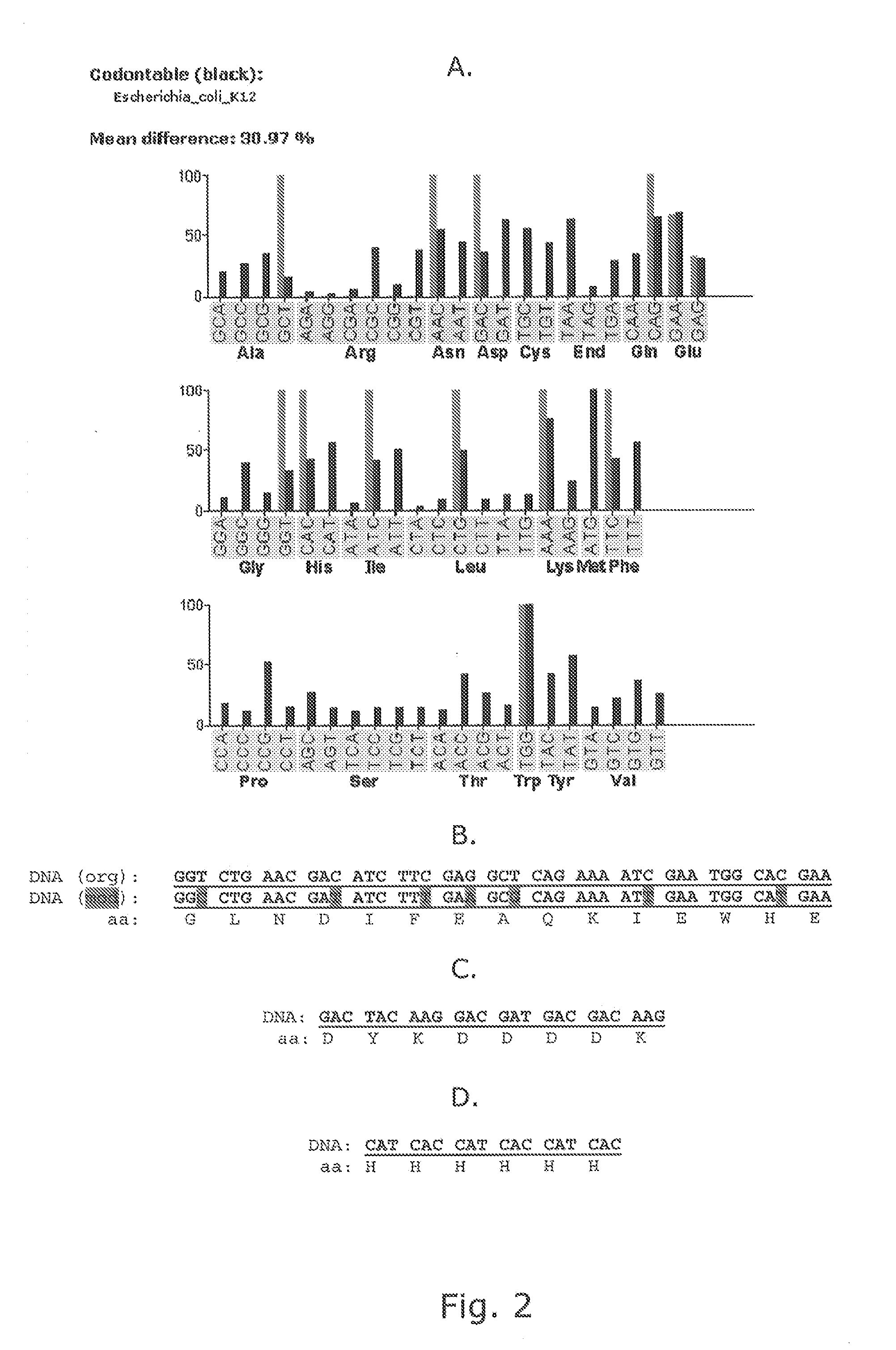
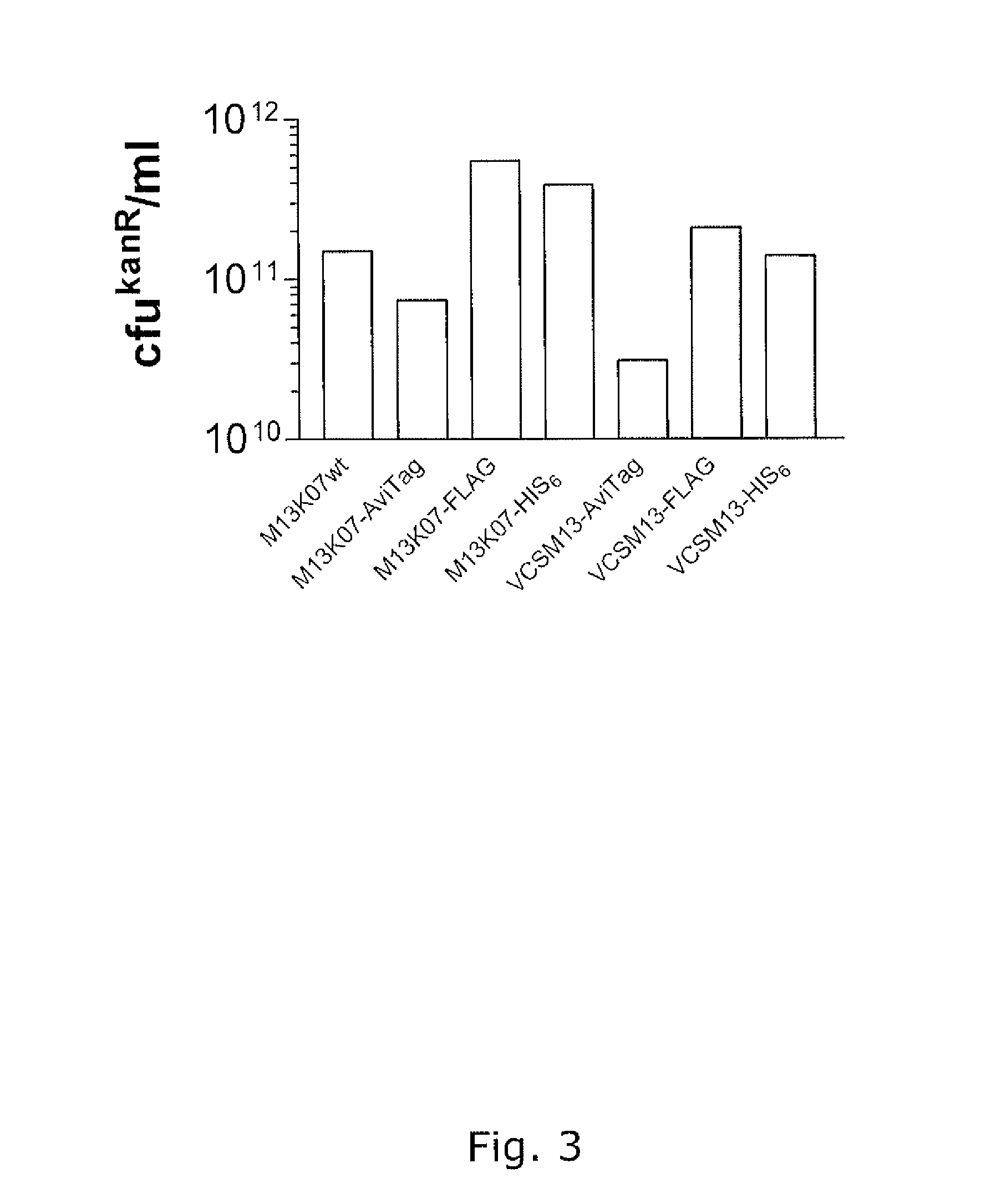
[0155]Modified helper phages M13K07 (SEQ ID NO: 31) and VCSM13 (SEQ ID NO: 32), with FLAG-pVII, HIS6-pVII, and AviTag-pVII may show a very broad potential for expanding the use of phage display technology, but it is of crucial importance that the fusion peptides do not compromise the functionality of the helper phage, thus titration of the phages are an important verification parameter. In addition the peptides fused to pVII must be accessible for the subsequent detection and / or immobilisation. This example support the fact that both pVII-modified helper phages can harbor a variety of peptides for detection and / or immobilization purposes and that these fusion peptides do not affect the infectivity of the phages.
[0156]Whilst early results from Endemann and Model (PMID: 7616570) indicated that the filamentous phage (Ff) capsid protein pVII did not tolerate exogenous fusions, it has later been shown that both phagemid-based (Gao et al (...
example 2
Functionality of Modified Helper Phages in Packaging of Phagemids
[0171]The promise of the invention is the use the modified helper phages for functional packaging of phagemids displaying a folded domain on a phage coat protein other than pVII, preferably in pIII or pVIII. The following examples support that modified helperphages with different peptides fused to pVII are able to perform functional phagemid packaging and that these phagemids display both functional pVII peptide fusion as well as functional folded domains fused to their pIII coatproteins. In this manner the examples also serve for bispecific display using phagemids.
Reagents
[0172]All media and buffers were prepared essentially as described in Sambrook et al (Molecular cloning: a laboratory manual (Cold Spring Harbor Laboratory Press)). The anti-M13-HRP antibody and the M2 and M5 antibodies were purchased from GE Healthcare Bio-Sciences AB (Uppsala, Sweden) and Sigma-Aldrich (Oslo, Norway), respectively, whereas the F23....
example 3
Genomic Phage Display on pIII and pVII
[0185]The invention allows for the generation of a genomic phage vector with display properties on pVII coatproteins. Such display will not affect the infectivity of virions like pIII display. Furthermore, the invention fosters bispecific display on pVII and pIII / pVIII, or even all three coat proteins simultaneously. The following example supports bispecific display on pIII and pVII in a genomic phage display system showing that the construct behave completely like wildtype phages with respect to propagation, virion assembly, viron concentration, pIII display phenotype and that it indeed is selectively in vivo biotinylated at the pVII peptide fusion.
Reagents
[0186]All media and buffers were prepared essentially as described in Sambrook et al (Molecular cloning: a laboratory manual (Cold Spring Harbor Laboratory Press)). The anti-M13-HRP antibody was purchased from GE Healthcare Bio-Sciences AB (Uppsala, Sweden) and the F23.2 and GB113 antibodies ...
PUM
 Abstract
Description
Claims
Application Information
 - R&D
- Intellectual Property
- Life Sciences
- Materials
- Tech Scout
- Unparalleled Data Quality
- Higher Quality Content
- 60% Fewer Hallucinations
Browse by: Latest US Patents, China's latest patents, Technical Efficacy Thesaurus, Application Domain, Technology Topic, Popular Technical Reports.
© 2025 PatSnap. All rights reserved.Legal|Privacy policy|Modern Slavery Act Transparency Statement|Sitemap|About US| Contact US: help@patsnap.com


