
Fusion Protein Inhibiting Osteoclast Formation, Preparation Method and Medicine Compositions Thereof

a technology of fusion protein and osteoclast, which is applied in the field of biotechnique for preparation of fusion protein, fusion protein and pharmaceutical composition, can solve the problems of osteoporosis that often leads to bone fracture, and the risk of endometrial carcinoma in patients is considerable, so as to inhibit osteoclast formation

Inactive Publication Date: 2011-10-06
SHANGHAI KEXIN BIOTECH
View PDF4 Cites 0 Cited by
  • Summary
  • Abstract
  • Description
  • Claims
  • Application Information

AI Technical Summary

Benefits of technology

[0009]The second goal of this invention is to provide fusion protein that can be used to inhibit osteoclast formation.

Problems solved by technology

Osteoporosis often leads to bone fracture and happens quite often in old people, especially women.
However, the application was declined twice in 2005 and 2006 because there were considerable chances of risk for the patients to have a carcinoma of the endometrium after the treatment.

Method used


Image

  • Fusion Protein Inhibiting Osteoclast Formation, Preparation Method and Medicine Compositions Thereof
  • Fusion Protein Inhibiting Osteoclast Formation, Preparation Method and Medicine Compositions Thereof
  • Fusion Protein Inhibiting Osteoclast Formation, Preparation Method and Medicine Compositions Thereof

Examples


example 1

Expression of Fusion Protein RIG

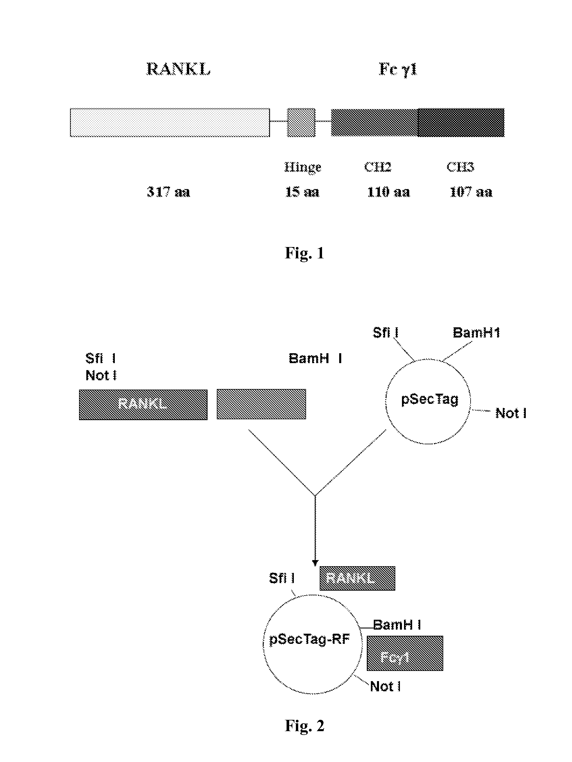
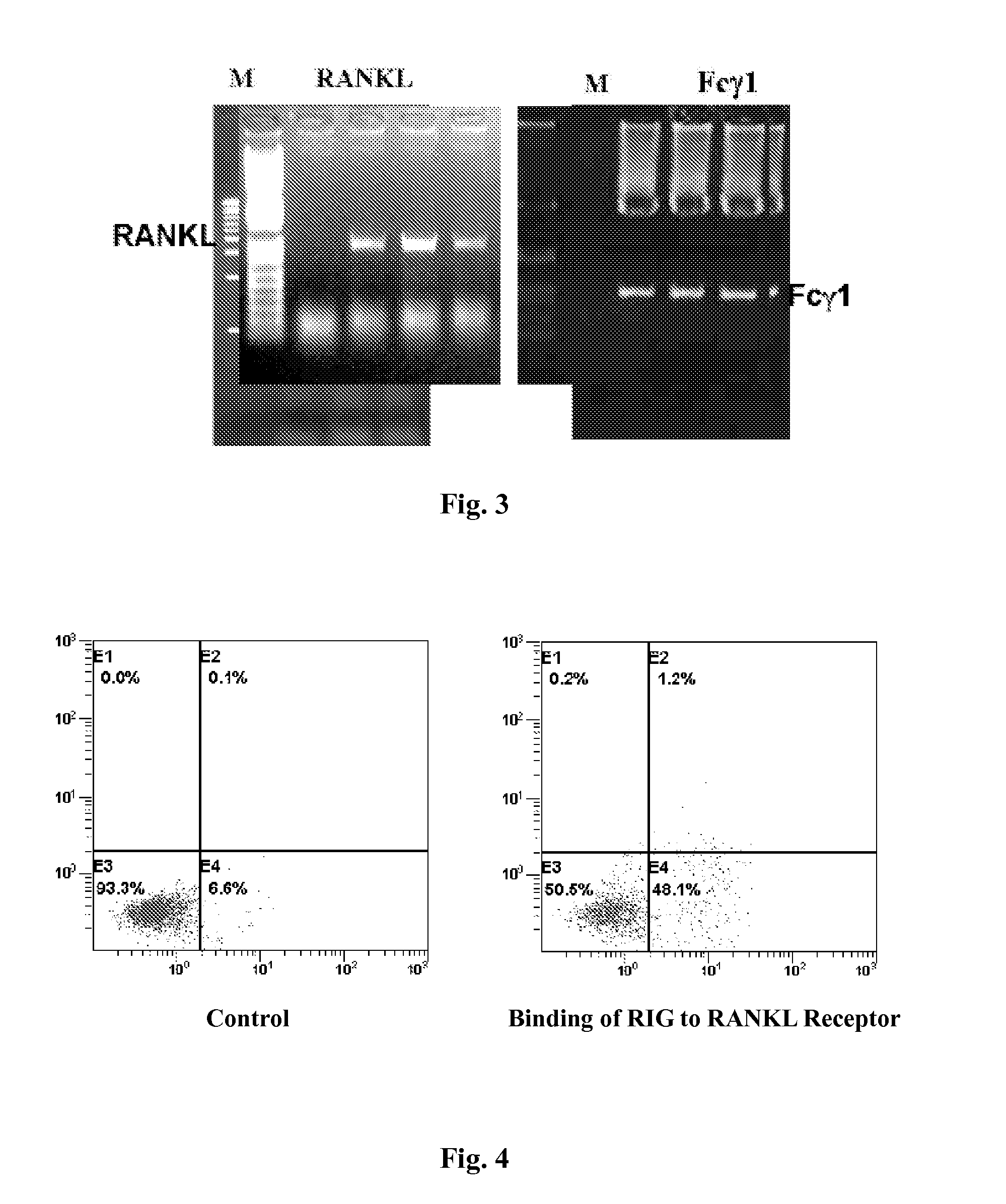
[0032]As illustrated in FIG. 2, the expression of the fusion protein includes the following steps: T lymphocytes and B lymphocytes were drawn from circumference blood and purified, from which mRNA were extracted. The specific primers were used to amplify the RANKL and Fcγ genes from T lymphocytes and B lymphocytes respectively using RT-PCR method. The primers used are from:

5′-GGCCCCGAGGGCCATGCGCCGCGCCAGCAGAGASEQ ID NO: 3C-3′; 5′-GGATCCGATCTATATCTCGAACTTTAAAAGC-3′;SEQ ID NO: 45′-GGATCCGAGCCCAAATCTTGTGAC-3′;SEQ ID NO: 55′-GCGGCCGCTCATTTACCCGGAGACAGGGAGASEQ ID NO: 6G-3′. 

[0033]The amplified RANKL and Fcγ genes were analyzed on 1% agarose electrophoresis. As shown in FIG. 3, the RANKL amplification product is about 970 bp long, while the Fcγ DNA amplification product is about 714 bp long. The two PCR products were cloned separately into CR4-TOPO vector (Invitrogen, CA) to obtain pRANKL and pFcγ plasmids respectively. The recombinant plasmids were sequence...

example 2

Binding Experiment of RIG and RANK (RANKL Receptor)

[0035]1*106 THP1 monocytes were grown in MCSF and RANKL medium for 72 hours and mixed with 5 μg purified RIG protein from Implementation 1 for additional 1 hour at 4° C. 5 μl FITC labeled anti human RANKL antibody (CALTAG, CA) was then added into the mixture and flow cytometry was used to examine FITC positive cells. As shown in FIG. 4, the result indicates that RIG protein bind to RANK which made the THP1 cell FITC positive. In FIG. 4, SSC-H presents the pellet number in cytoplasm, FSC-H presents the cell size, FL2-H presents PE and FL1-H presents FITC.

example 3

Binding Experiment of RIG and FcγRII Receptor

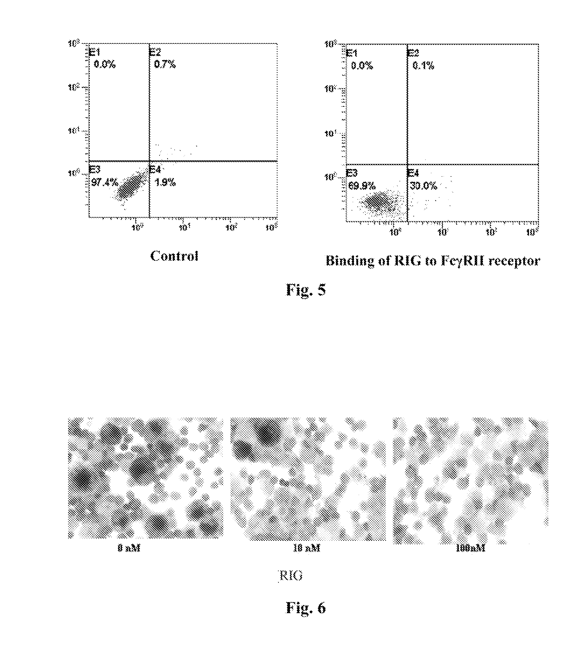
[0036]1*106 HMC-1 cells were mixed with 5 μg purified RIG protein from Implementation 1 for 1 hour at 4° C. 5 μl FITC labeled anti human IgG antibody (CALTAG, CA) was then added into the mixture and flow cytometry was used to examine FITC positive cells. As shown in FIG. 5, the result indicates that RIG protein can bind to FcγRII receptor which made the HMC-1 cell FITC positive. In FIG. 5, SSC-H presents the pellet number in cytoplasm, FSC-H presents the cell size, FL2-H presents PE and FL1-H presents FITC.


PUM

PropertyMeasurementUnit
diameteraaaaaaaaaa
diameteraaaaaaaaaa
nucleic acid sequenceaaaaaaaaaa

Abstract

The present invention provides a coding gene having the nucleic acid sequence shown as SEQ ID NO:1 and the fusion protein RIG (SEQ ID NO:2) that inhibits osteoclast formation. The present invention also provides the preparation method for the fusion protein RIG as well as synthetic oligo-nucleotide primers, plasmids and host cells used in the method and a medicine having the above fusion protein RIG as active ingredient. The fusion protein RIG in present invention is derived from humanized immunoglobulin and RANKL with a flexible hinge region. RIG can cross link the cell surface receptor RANK and Fcγ 1 to induce a cytosolic inhibitory signal leading to the inhibition of osteoclast formation. The fusion protein RIG in present invention can play an essential role in treating osteoporosis and bone resorption diseases caused by tumor metastasis.

Description

FIELD OF THE INVENTION[0001]The present invention relates to biomedical fields. In particular, the present invention relates to a bio-technique for preparation of a fusion protein. More particularly, the present invention relates to a fusion protein and pharmaceutical compositions that can be used to treat osteoporosis and bone resorption caused by tumor metastasis.BACKGROUND OF THE INVENTION[0002]Bone formation and bone resorption are two highly related and balanced processes involved in normal bone remodeling. For osteoporosis patients, their osteoclast activity is elevated to such an extent that bone absorption rate exceeds bone formation rate. This process causes bone disease without the loss of bone mineral. Bone is also a frequent target for invasion and metastasis of tumors such as multiple myeloma, mammary cancer, prostatic cancer and lung cancer. During the metastasis process, cancer cells interact with the osteoclast precursors in bone marrow to differentiate into osteocla...

Claims


Application Information

Patent Timeline
Patent Type & Authority Applications(United States)
IPC IPC(8): A61K39/395C07H21/04C07K19/00C12P21/02A61P19/08A61P19/10
CPCC07K14/70575A61K38/00C07K2319/30A61P19/08A61P19/10A61P35/00
Inventor BAO, JUNCHEN, HAIMINGZHU, DAOCHENG
Owner SHANGHAI KEXIN BIOTECH
Learn More