
Apparatus and method for generating stereoscopic viewing image based on three-dimensional medical image, and a computer readable recording medium on which is recorded a program for the same

a three-dimensional medical image and stereoscopic technology, applied in diagnostic recording/measuring, digital output to print units, instruments, etc., can solve the problems of not all the apparatuses for outputting medical images support nor are required to support stereoscopic display

Inactive Publication Date: 2011-09-29
FUJIFILM CORP
View PDF15 Cites 39 Cited by
  • Summary
  • Abstract
  • Description
  • Claims
  • Application Information

AI Technical Summary

Benefits of technology

The present invention relates to a stereoscopic viewing image generation apparatus and method that can generate a stereoscopic viewing image and a non-stereoscopic viewing image based on a three-dimensional medical image and an image generation condition. The apparatus and method can selectively output either the stereoscopic viewing image or the non-stereoscopic viewing image based on the output image selection condition. The technical effect of this invention is to provide a more efficient and flexible tool for generating stereoscopic viewing images that can be used in various applications such as medical diagnosis and treatment planning.

Problems solved by technology

Therefore, if an area is specified for one of the parallax images, as the method described in U.S. Pat. No. 7,572,009, unpleasant sensation may be felt when specifying the area or the determined area may differ from the desired area of the stereoscopically displayed image due to the difference in direction between the actual visual line for viewing the stereoscopically displayed image and the visual line of the parallax image for which the area is specified.
Further, in the method described in U.S. Pat. No. 7,572,009, images serving as the basis of stereoscopic display are only two images obtained by imaging, and another image can not be used for the area specification.
But, in actual clinical practices, not all of the apparatuses for outputting medical images support nor are required to support stereoscopic display.

Method used


Image

  • Apparatus and method for generating stereoscopic viewing image based on three-dimensional medical image, and a computer readable recording medium on which is recorded a program for the same
  • Apparatus and method for generating stereoscopic viewing image based on three-dimensional medical image, and a computer readable recording medium on which is recorded a program for the same
  • Apparatus and method for generating stereoscopic viewing image based on three-dimensional medical image, and a computer readable recording medium on which is recorded a program for the same

Examples


first embodiment

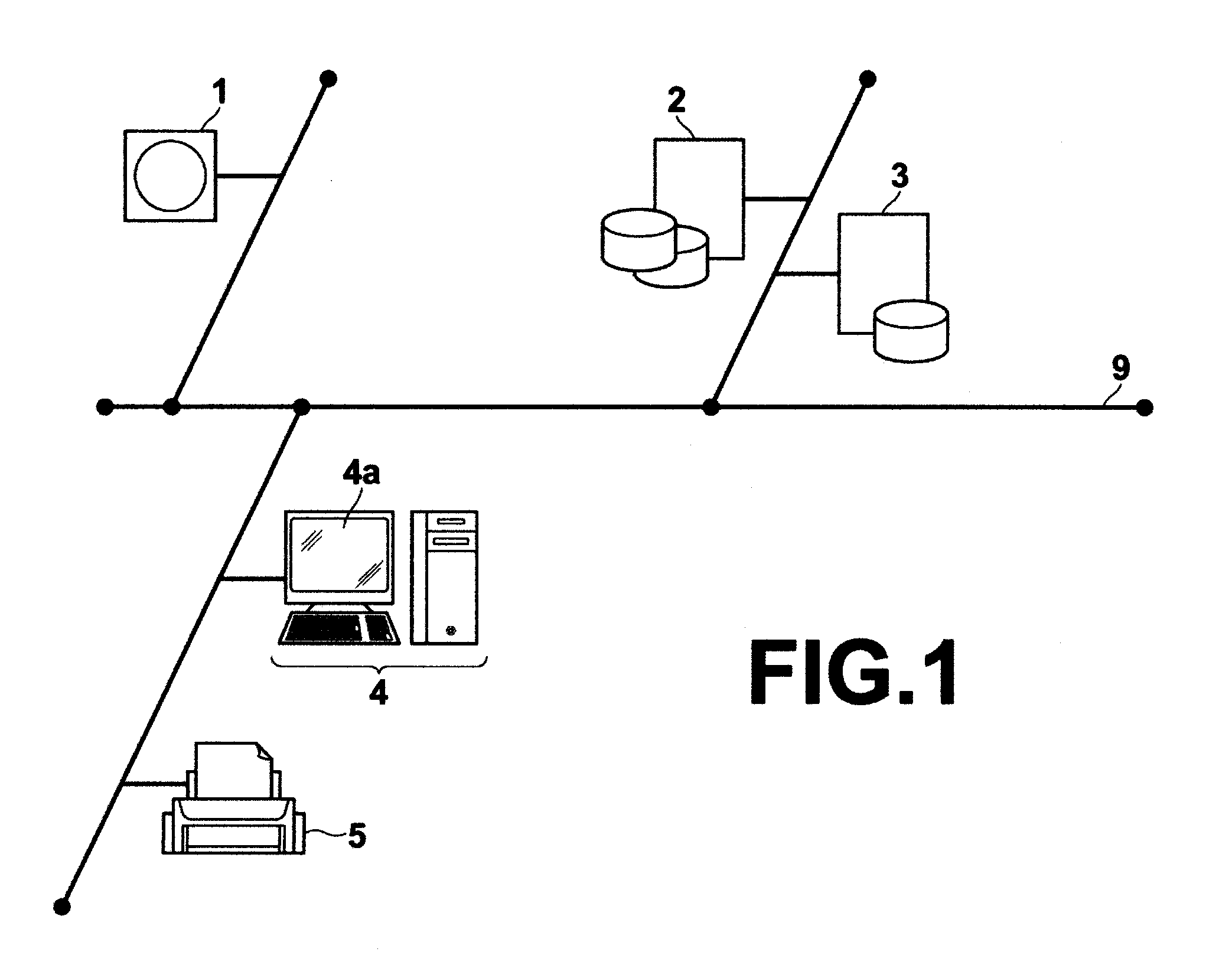
[0069]FIG. 3 is a flowchart illustrating a flow of user operation, calculation processing, display processing, and the like performed under the execution of stereoscopic viewing image generation software according to the present invention. First, image processing workstation 4 obtains image data of a processing target three-dimensional medical image V from image storage server 2 through image retrieval and acquisition processing of a known image retrieval system or of a known ordering system (#1).

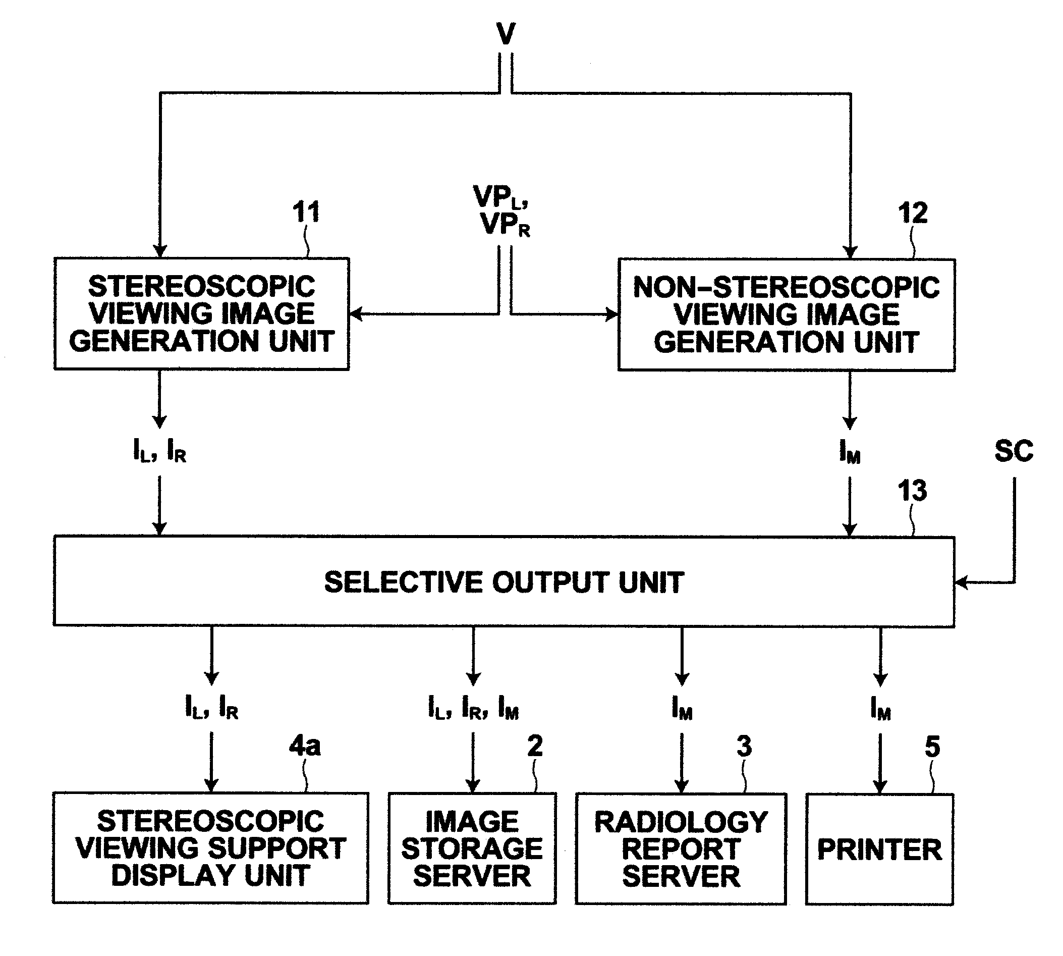
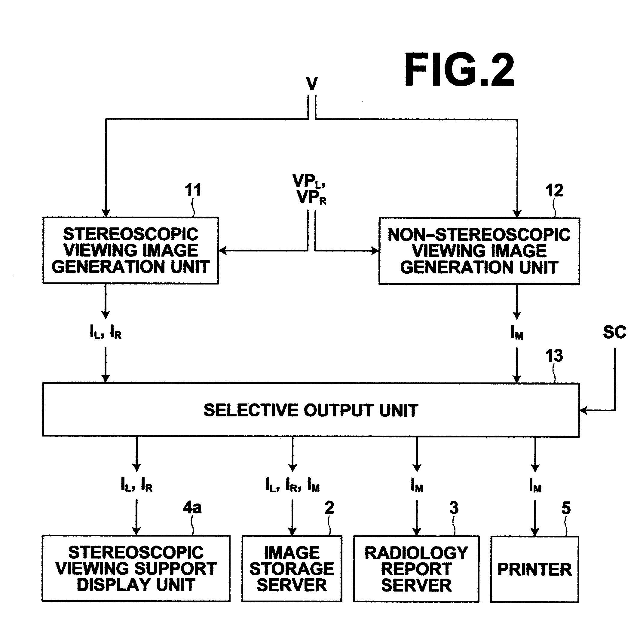
[0070]Then, stereoscopic viewing image generation unit 11 generates left eye parallax image IL and right eye parallax image IR for implementing a stereoscopic output based on the three-dimensional medical image V, left eye viewpoint position VPL, and right eye viewpoint position VPR (#2), and non-stereoscopic viewing image generation unit 12 determines one viewpoint equivalent to the stereoscopic output from the left eye viewpoint position VPL and right eye viewpoint position VPR and genera...

second embodiment

[0090]Next, an embodiment in which switching between a stereoscopic display and a non-stereoscopic display is performed on stereoscopic viewing support display unit 4a connected to image processing workstation 4 in the medical image diagnosis system shown in FIG. 1 will be described, as the present invention.

[0091]FIG. 8 is a block diagram illustrating a portion of the function of image processing workstation 4 relevant to the stereoscopic viewing image generation processing according to the second embodiment of the present invention. As shown in FIG. 8, stereoscopic viewing image generation processing of the second embodiment of the present invention is achieved realized by stereoscopic viewing image generation unit 11, non-stereoscopic viewing image generation unit 12, cross-sectional image generation unit 14, and display control unit 15. In FIG. 8, the three-dimensional medical image V, left eye viewpoint position VPL, right eye viewpoint position VPR, left eye parallax image IL,...


PUM


Abstract

A stereoscopic viewing image generation unit generates, using a three-dimensional medical image representing a subject as input, a stereoscopic viewing image for stereoscopic output of the subject based on a given image generation condition, and a non-stereoscopic viewing image generation unit generates a non-stereoscopic viewing image for non-stereoscopic output equivalent to the stereoscopic output based on the three-dimensional medical image and the image generation condition of the stereoscopic viewing image.

Description

BACKGROUND OF THE INVENTION[0001]1. Field of the Invention[0002]The present invention relates to an apparatus and method for generating a stereoscopic viewing image from a three-dimensional medical image, and a computer readable recording medium on which is recorded a program for the same.[0003]2. Description of the Related Art[0004]Technologies for performing a stereoscopic display are known.[0005]For example, a stereoscopic display apparatus for a tomographic image in which two parallax images of a cross-section of a test object viewed from left and right eyes are generated based on a plurality of tomographic images obtained by CT or the like and generated parallax images are displayed on two displays is proposed as described, for example, in Japanese Unexamined Patent Publication No. 62 (1987)-016741. Here, the parallax images can be obtained, based on each of left and right viewpoints, by adding up a grey value of each pixel of tomographic images on a visual line connecting betw...

Claims


Application Information

Patent Timeline
Patent Type & Authority Applications(United States)
IPC IPC(8): G06F3/12G06T15/00
CPCG06T7/0022H04N13/0029G06T15/00H04N13/0454H04N13/0456H04N13/004A61B5/742G06T7/97H04N13/139H04N13/156H04N13/359H04N13/361
Inventor SAKURAGI, FUTOSHI
Owner FUJIFILM CORP
Learn More