
Biopsy needle with vacuum assist

a technology of biopsy needle and vacuum assist, which is applied in the field of surgical devices for biopsy sampling of tissue, can solve the problems of longer healing/recovery time and greater discomfort for patients

Inactive Publication Date: 2011-06-23
COOK INC
View PDF10 Cites 136 Cited by
  • Summary
  • Abstract
  • Description
  • Claims
  • Application Information

AI Technical Summary

Benefits of technology

"The patent describes a surgical cutting instrument that can be used to procure tissue samples. The instrument includes a cannula with a distal end shaped to cut tissue, and a stylet that can be moved from a retracted position to a cutting position. The stylet has a sharp distal end and a sample collection region that is in fluid communication with a vacuum port. The instrument also includes a control handle with a vacuum source and a sealing sleeve that can be moved from a sealed position to a released position. The sealing sleeve prevents fluid communication between the vacuum source and the lumen of the stylet when in the sealed position, while the released position allows fluid flow. The instrument can facilitate the procurement of tissue samples and is easy to use."

Problems solved by technology

However, because biopsy is an invasive procedure that involves insertion through the skin and the removal of tissue, the larger the needle used to procure the tissue sample, the greater the discomfort to the patient and longer the healing / recovery time.

Method used


Image

  • Biopsy needle with vacuum assist
  • Biopsy needle with vacuum assist
  • Biopsy needle with vacuum assist

Examples


Embodiment Construction

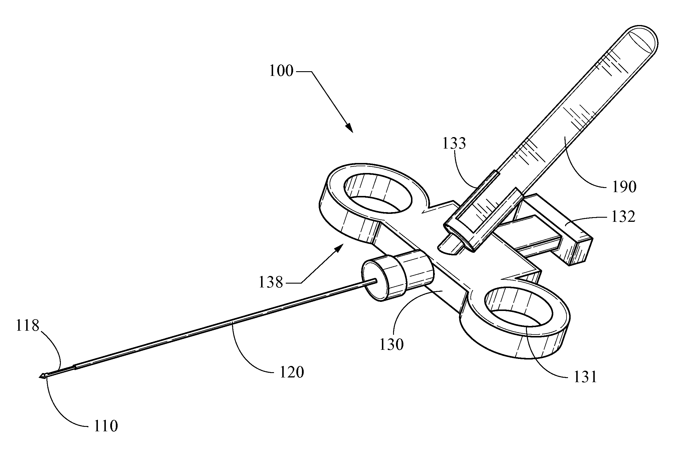
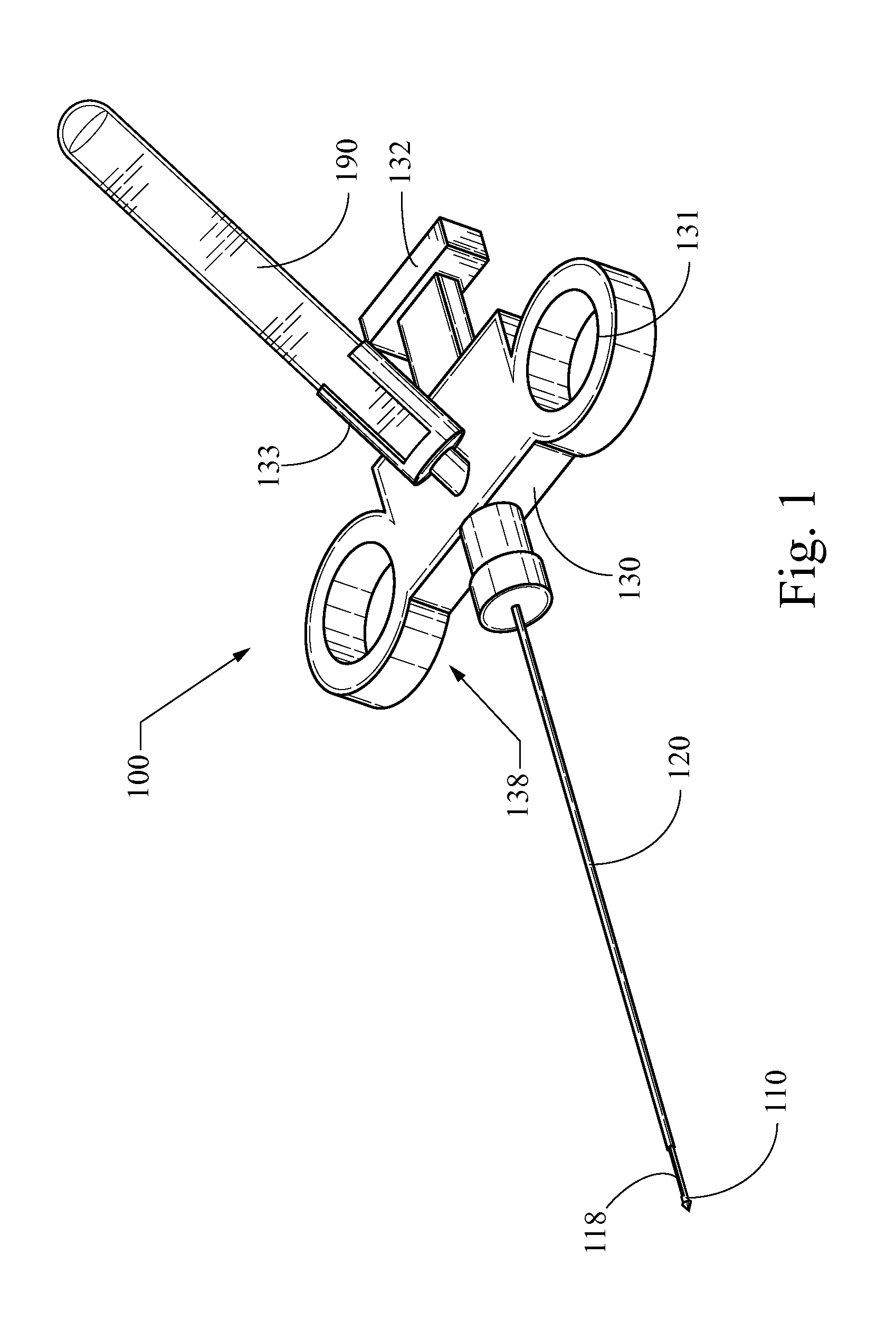
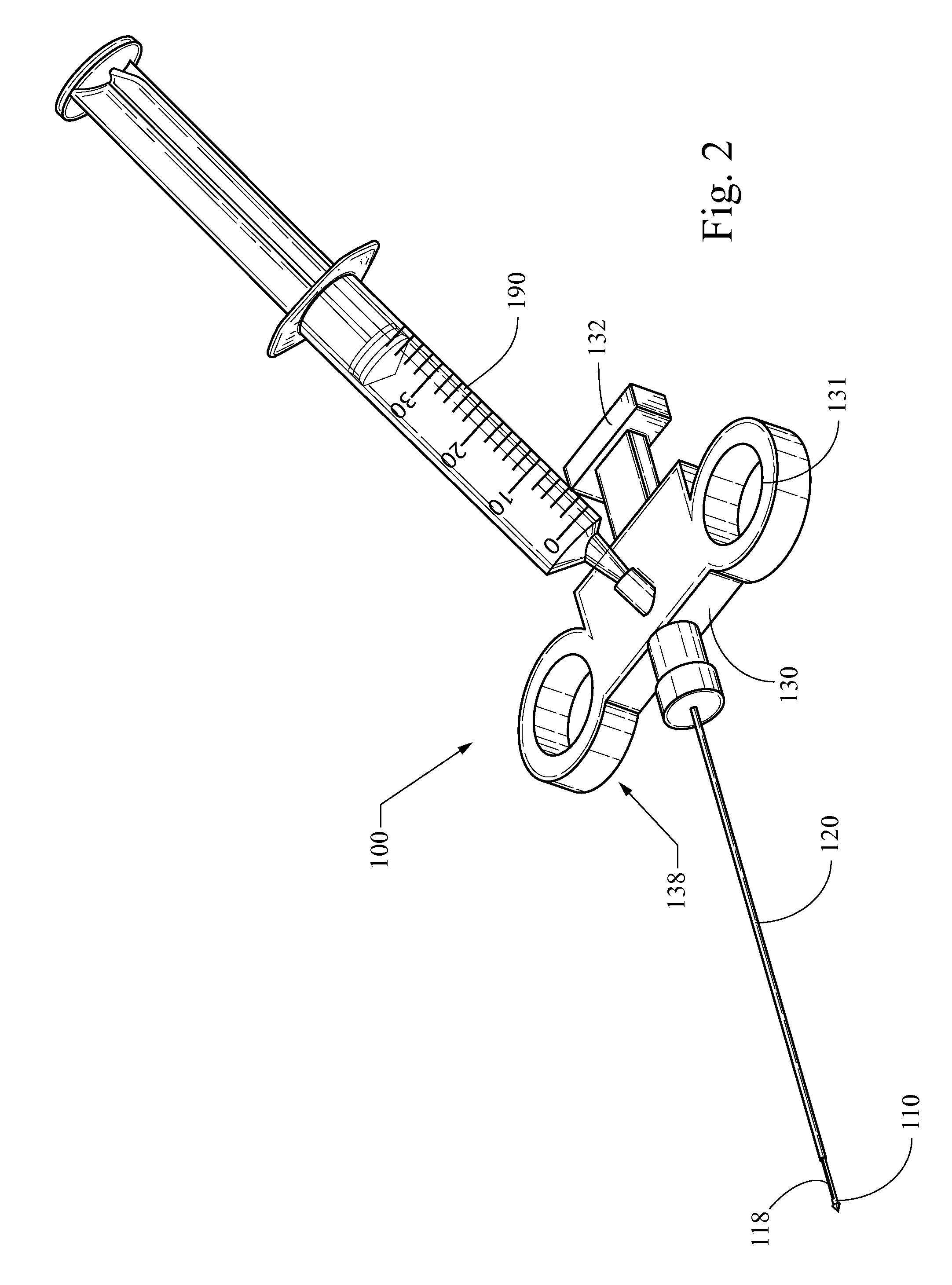
Referring now to the figures, FIGS. 1-3 illustrate embodiments of a surgical cutting instrument for biopsy sampling of tissue. More specifically, FIGS. 1 and 2 illustrate embodiments of a surgical cutting instrument 100 having a releasably attachable and replaceable fixed volume vacuum source 190, while FIG. 3 illustrates an embodiment having an integrally formed fixed volume vacuum source 190. Note that throughout this specification, like reference numbers refer to like elements in the Figures.

As shown in the embodiments of FIGS. 1 and 2, the surgical cutting device includes a control handle 130 connected to an elongated tube or cannula 120, a tissue penetrating stylet 110, and a fixed volume vacuum source 190. The control handle 130 may include a body 138 having two finger holes 131 disposed on opposite sides thereof. An attachment member 133 configured to releasably engage and attach the fixed volume vacuum source 190 to the control handle 130 may be disposed on the external surf...


PUM


Abstract

A surgical cutting device comprises a cannula attached to an actuation mechanism that moves the cannula from a cocked position to a cutting position, a stylet including a distal portion having a sharp distal end, a sample collection region, a vacuum port, and a lumen extending from the vacuum port to the sample collection region such that the sample collection region is in fluid communication with the first vacuum port. When the cannula is in the cocked position, the sample collection region of the stylet is disposed distal of the cannula. The device also includes a fixed volume vacuum source. When cannula is in the cocked position, the vacuum port is not in fluid communication with the fixed volume vacuum source, and when said cannula is in the cutting position, the vacuum port is in fluid communication with the fixed volume vacuum source.

Description

BACKGROUNDThe present invention relates generally to medical devices and more particularly to a surgical device for biopsy sampling of tissue.Biopsy is the removal and study of body tissue for medical diagnosis. Typically, physicians obtain biopsy samples in order to detect abnormalities, such as cancer, and to determine the extent to which cancerous tissue has spread. Generally, tissue samples are acquired from different areas of the body using biopsy instruments. Common biopsy instruments comprise a two-part needle assembly, commonly referred to as a stylet and cannula, operated by a spring-loaded handle of the type disclosed in U.S. Pat. No. 5,538,010, the entirety of which is incorporated herein by reference. In use, the biopsy device is inserted through the skin to the target biopsy site with the cannula in the “cocked,” or retracted position, and the stylet in the retracted position. The stylet is then advanced out of the cannula to its deployed position, which is distal of th...

Claims


Application Information

Patent Timeline
Patent Type & Authority Applications(United States)
IPC IPC(8): A61B10/02
CPCA61B10/0275A61B2010/0208A61B10/0283
Inventor DELAP, DENNIS J.KOEHLER, CLEVE S.CHILDRESS, ROBERT S.MYERS, RANDY JOE
Owner COOK INC
Learn More