
Device, system and method for multiple core biopsy

a biopsy and multiple core technology, applied in the field of biopsy devices, can solve the problems of unattainable histopathological assessment, a large amount of specimen crushed along the outer edges, and a time-consuming and labor-intensive process for standard tissue sampling

Inactive Publication Date: 2011-05-26
CRUX MEDICAL INNOVATIONS INC
View PDF51 Cites 43 Cited by
  • Summary
  • Abstract
  • Description
  • Claims
  • Application Information

AI Technical Summary

Problems solved by technology

This is a tedious and time consuming process for standard tissue sampling and even more so for surveillance biopsies where in excess of 50 specimens may be taken.
The traditional biopsy forceps design creates some of the complications experienced today but is accepted as the standard as there is no better alternative available.
However when this is accomplished much of the specimen is crushed along the outer edges.
As much as 50% of the specimens are suboptimal or inadequate showing crush artifact or they are superficial so that histopathological assessment is unattainable.
Unfortunately even with these larger cups a great percentage is not adequate due to shearing or crush artifact.
These inadequate samples have held the key to a more accurate diagnosis but due to their condition were not salvageable.
Research indicates this product has not been very successful.
Its design actually limits the size of tissue sample and causes additional crushing which both reduce the quality for pathology.
The conclusion is that physicians are not willing to give up quality of pathology to save time.
This is a very tedious, time consuming process with plenty of opportunity to further damage the specimen.

Method used


Image

  • Device, system and method for multiple core biopsy
  • Device, system and method for multiple core biopsy
  • Device, system and method for multiple core biopsy

Examples


Embodiment Construction

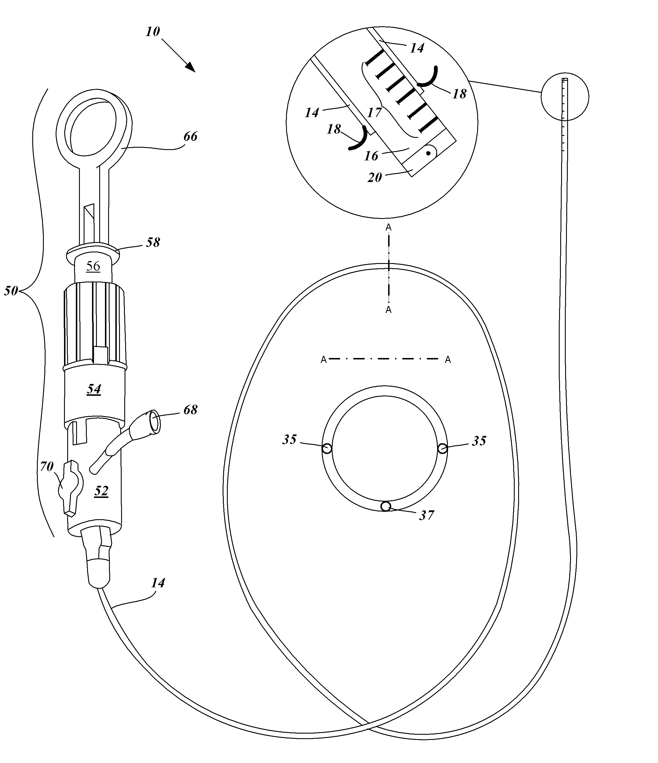
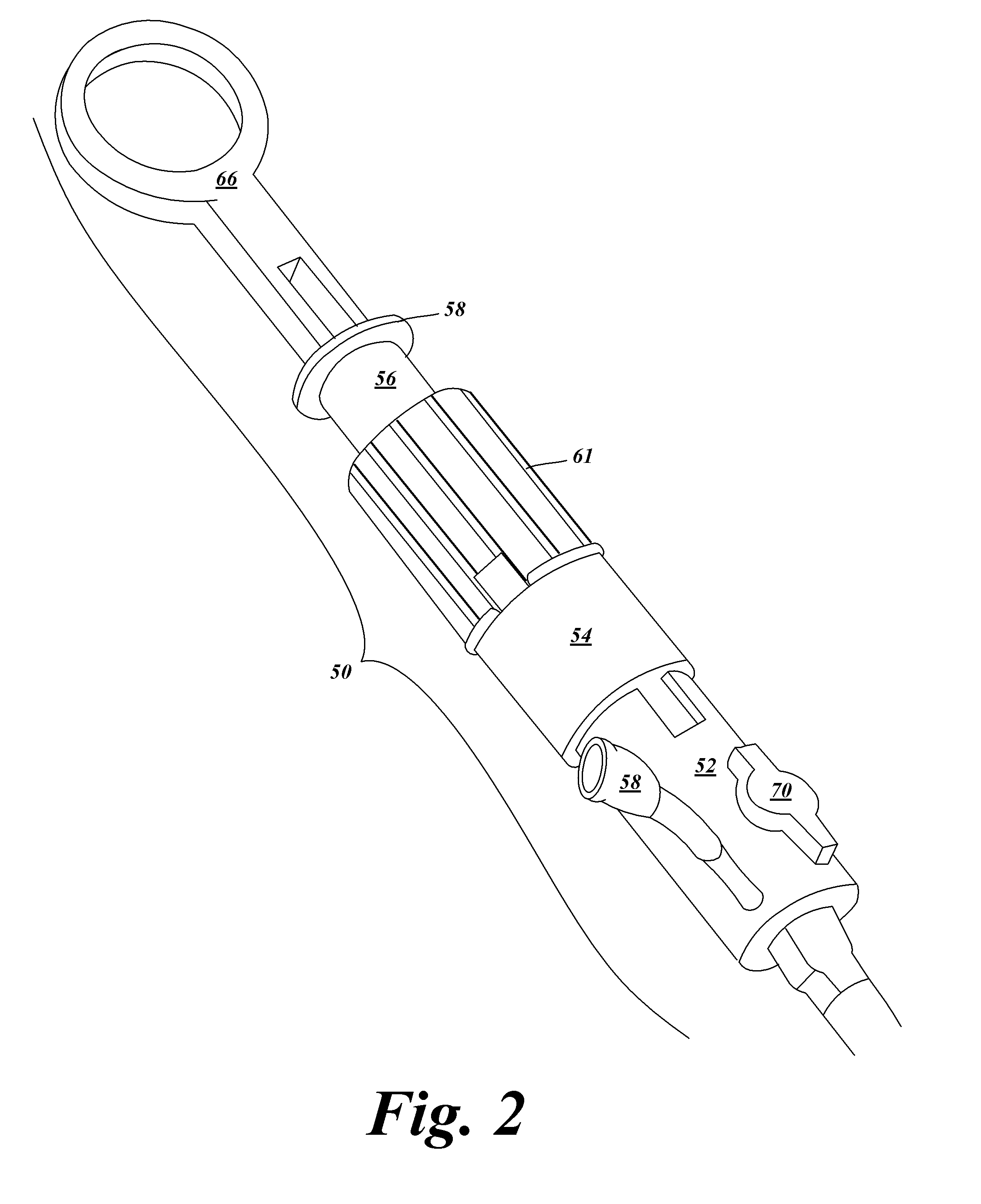
[0046]A Multiple Core Biopsy (MCB) device with a substantially cylindrically-shaped core and a sharp forward-facing cutting edge is configured to acquire multiple tissue biopsy samples during a single endoscopic procedure. The tissue biopsy sections are either temporarily stored within the specimen chamber, or aspirated through the device into a specimen management system maintaining sampling order and specimen orientation. If they are temporarily stored in the specimen chamber the specimens are removed by expelling into a specimen management system after the MCB is removed for the scope. Expulsion is achieved by a reverse fluid flush in communication with the specimen chamber with preservation of the specimen's order and orientation into a specimen management system. Once the samples are contained in the specimen management system it is removed and labeled for processing. With the specimen chamber cleared the MCB is ready for alternate site sampling. The MCB device specimen chamber...


PUM


Abstract

A core biopsy device having a forward-facing cutting edge with either single or dual counter rotating blades is configured to acquire multiple biopsy specimens and temporarily store them within a specimen chamber located beneath the cutting edge. The core biopsy device is insertable and removable from a biopsy channel of an endoscope. The core biopsy device includes an endoscopically visible ruler with depth gauge increments. Other embodiments include deployable depth stops to restrain the plunging of the specimen chamber to not exceed distances preset or selected by the user. Multiple samples contained within the specimen chamber are removed via flushing from a syringe adapter when the device is removed from the endoscope. Alternatively the multiple specimens are removed under applied suction to an external chamber located outside the patient. The order and orientation of the removed specimens are preserved.

Description

CROSS REFERENCES TO RELATED APPLICATIONS[0001]This application claims the benefit of priority to and incorporates by reference in its entirety U.S. Provisional Patent Application Ser. No. 61 / 264,628 filed Nov. 25, 2009.FIELD OF THE INVENTION[0002]An embodiment of the invention relates generally to biopsy devices used in endoscopic and other tissue sampling procedures.BACKGROUND OF THE INVENTION[0003]The traditional biopsy forceps used today typically gather just one biopsy specimen at a time. The forceps must be removed from the scope each time to retrieve the single specimen. This is a tedious and time consuming process for standard tissue sampling and even more so for surveillance biopsies where in excess of 50 specimens may be taken. A large variety of endoscopic biopsy forceps are commercially available today with the majority of the market converting to disposable forceps over the last 15 years. The current design has not fundamentally changed since its inception over 30 years ...

Claims


Application Information

Patent Timeline
IPC IPC(8): A61B10/02
CPCA61B10/0266A61B10/0283A61B10/04A61B2019/462A61B2010/0093A61B2010/0225A61B2019/303A61B10/06A61B2090/033A61B2090/062
Inventor CLEMENTS, ROBERTFACE, ANDREW R.
Owner CRUX MEDICAL INNOVATIONS INC
Learn More