
Method and System for Detection 3D Spinal Geometry Using Iterated Marginal Space Learning

Inactive Publication Date: 2011-03-17
SIEMENS HEALTHCARE GMBH
View PDF6 Cites 29 Cited by
  • Summary
  • Abstract
  • Description
  • Claims
  • Application Information

AI Technical Summary

Benefits of technology

[0005]The present invention provides a method and apparatus for automatic detection of spinal geometry in 3D images. Embodiments of the present invention combine efficient local object detection based on marginal space learning (MSL) with a global probabilistic model that incorporates pose priors on the nine dimensional parameter spaces that encode the position, orientation, and scale of the individual intervertebral disks. Embodiments of the present invention utilize a database-guided detection paradigm and can thus be easily trained for spine detection in computed tomography (CT) and magnetic resonance (MR) images acquired with different sequences.

Problems solved by technology

An automatic procedure for extracting the spinal geometry faces various challenges, however.
Varying contrasts and image artifacts can compromise the detection of intervertebral disks based on local image features.

Method used


Image

  • Method and System for Detection 3D Spinal Geometry Using Iterated Marginal Space Learning
  • Method and System for Detection 3D Spinal Geometry Using Iterated Marginal Space Learning
  • Method and System for Detection 3D Spinal Geometry Using Iterated Marginal Space Learning

Examples


Embodiment Construction

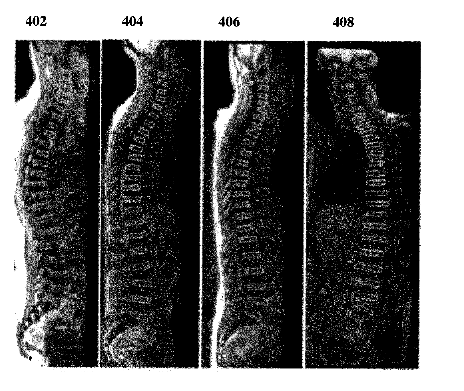
[0016]The present invention is directed to a method and apparatus for detecting 3D spinal geometry object in medical images, such as computed tomography (CT) or magnetic resonance (MR) images. Embodiments of the present invention are described herein to give a visual understanding of the anatomical object detection method. A digital image is often composed of digital representations of one or more objects (or shapes). The digital representation of an object is often described herein in terms of identifying and manipulating the objects. Such manipulations are virtual manipulations accomplished in the memory or other circuitry / hardware of a computer system. Accordingly, is to be understood that embodiments of the present invention may be performed within a computer system using data stored within the computer system.

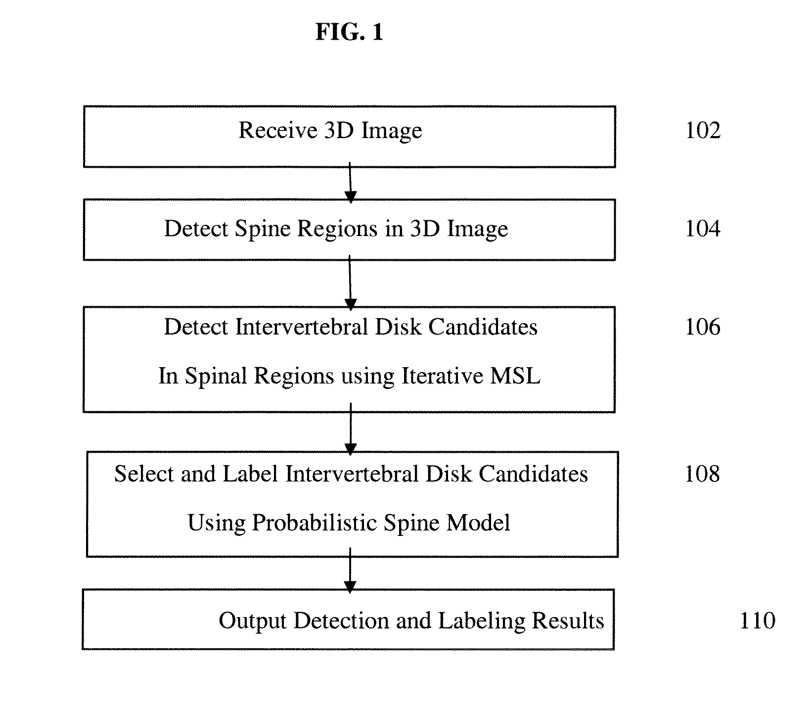
[0017]FIG. 1 illustrates a method for automatically detecting and labeling intervertebral disks in a 3D image according to an embodiment of the present invention. The meth...


PUM


Abstract

A method and apparatus for automatic detection and labeling of 3D spinal geometry is disclosed. Cervical, thoracic, and lumbar spine regions are detected in a 3D image. Intervertebral disk candidates are detected in each of the spine regions using iterative marginal space learning (MSL). Using a global probabilistic spine model, a separate one of the intervertebral disk candidates is selected for each of a plurality of labeled intervertebral disk locations.

Description

[0001]This application claims the benefit of U.S. Provisional Application No. 61 / 243,313, filed Sep. 17, 2009, the disclosure of which is herein incorporated by reference.BACKGROUND OF THE INVENTION[0002]The present invention relates to detection of 3D spinal geometry in images, and more particularly, to automated detection and labeling of 3D spinal disks in medical images using iterated marginal space learning.[0003]Examinations of the vertebral column with both Magnetic Resonance (MR) and Computer Tomography (CT) require a standardized alignment of the scan geometry with the spine. While in MR, the intervertebral disks can be used to align slice groups to position saturated bands, in CT the reconstruction planes need to be aligned. In addition to the position and orientation of the disks, physicians are typically interested in labeling the disks (e.g., C2 / C3, C5 / T1, L1 / L2 . . . ). Labeling the intervertebral disks allows one to quickly determine the anatomical location without err...

Claims


Application Information

Patent Timeline
IPC IPC(8): G06K9/00
CPCG06K2209/055G06T7/0087G06T2207/30012G06T2207/20081G06T2207/20076G06T7/143G06V2201/033
Inventor KELM, MICHAELZHOU, SHAOHUA KEVINZHENG, YEFENGSUEHLING, MICHAEL
Owner SIEMENS HEALTHCARE GMBH
Learn More