
Method for Decreasing the Size and/or Changing the Shape of Pelvic Tissues

a technology of pelvis and pelvis, applied in the field of surgical methods, can solve the problems of compromising sexual function, sexual dysfunction, and many women's unhappy with the size, shape and/or contour of the vagina or labia, and achieve the effect of not leading to loss of function or poor aesthetic results

Active Publication Date: 2010-01-07
ZIPPER RALPH
View PDF58 Cites 11 Cited by
  • Summary
  • Abstract
  • Description
  • Claims
  • Application Information

AI Technical Summary

Benefits of technology

[0013]Accordingly, it is the object of the present invention to provide a method to contour one or more pelvic tissues without leading to loss of function or poor aesthetic results. Additionally, one or more of the embodiments of the present invention may also be used to treat urinary incontinence.

Problems solved by technology

Many women are unhappy with the size, shape, and / or contour of the vagina or labia.
This enlargement and / or unsatisfactory shape or contour may lead to sexual dysfunction which may be anatomic or psychological in nature.
Although these surgeries may alter the size and shape of the vagina and labia, they may often compromise sexual function or create less than optimal aesthetic results.
The sexual dysfunction created by such surgeries may be secondary to stenosis of the vagina, shortening of the vagina, injury to muscles or nerves leading to pain or anesthesia, injury of the Graffenberg Spot, removal of the Graffenberg spot, or poor aesthetic appearance leading to psychological sexual dysfunction.
Injuries to the supporting structures of the vagina and surrounding tissues may also cause urinary incontinence.
Present treatments for urinary incontinence do not restore normal anatomic structure.

Method used


Image

  • Method for Decreasing the Size and/or Changing the Shape of Pelvic Tissues
  • Method for Decreasing the Size and/or Changing the Shape of Pelvic Tissues
  • Method for Decreasing the Size and/or Changing the Shape of Pelvic Tissues

Examples


first embodiment

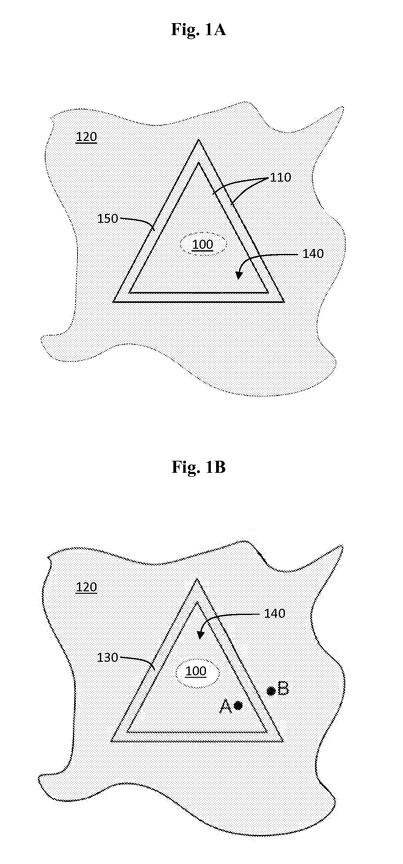
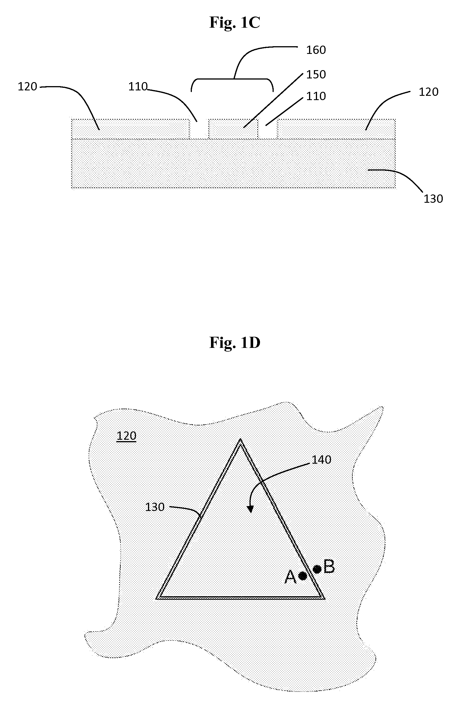
[0033]A first embodiment, depicted in FIGS. 1A-1D, may provide for protection of the Graffenberg Spot (G-Spot). In this embodiment, the vaginal mucosa of the G-Spot 100 may be left intact. At least one incision 110 of any known shape, preferably triangular-shaped, may be made around the G-Spot 100, as shown in FIG. 1A. The at least one incision 110 may be carried through the thickness of the vaginal mucosa 120. The at least one incision 110 may spare the endopelvic fascia 130. The at least one incision 110 may assume any known shape thereby defining the shape of an island 140 of tissue. In a preferred embodiment, as shown in FIGS. 1A-1D, a triangular-shaped island 140 of mucosa 120 may be created by the at least one incision 110. A strip 150 of mucosa 120 may be removed from the circumference of the island 140 to expose a channel 160 of endopelvic fascia 130, as shown in FIG. 1B and FIG. 1C. The diameter of this channel 160 will determine the final shape and / or size of the vagina. A...

second embodiment

[0034]A second embodiment, depicted in FIGS. 2A-2C, may provide for vaginal shaping without removal of fascia. In this embodiment, as shown in FIG. 2A, strips 250 of vaginal mucosa layer 220 may be removed while sparing the underlying endopelvic fascia 230 and nerve injury (see FIG. 2B). Rather than pulling the mucosal 220 edges together and creating a submucosal deformity, RF energy may be applied to shrink the endopelvic fascia 230 and bring the mucosal 220 edges closer together, as shown by the relative movement of point A and point B in FIGS. 2B and 2C. The limited penetration of RF energy acts to spare the underlying nerve structure and improves the thickness of underlying tissue. The mucosal 220 edges may be left “as is”, approximated with sutures or glue, or closed by any other manner known within the art. Although RF is the preferred energy source, any other types of energy known within the art including but not limited to laser energy, microwave energy, chemical energy, and...

third embodiment

[0035]A third embodiment, depicted in FIGS. 3A-3C, may provide for vaginal shaping without removal of mucosa. As shown in FIG. 3A, one or more incisions 310 may be made in the mucosa 320. The endopelvic fascia 330 or other submucosal tissue may be left attached to the mucosa 320. As shown in FIG. 3B, RF energy may be applied to the endopelvic fascia 330 or other submucosal tissue exposed between the one or more incision 310 margins. Such an application of energy will cause shrinkage of such endopelvic fascia 330 tissue with proportional contraction of the overlying mucosa 320 and spare the deep nerves and subfascial or subcutaneous tissue 335. Any such endopelvic fascia 330 that is left exposed (as expressly disclosed in all embodiments) may be treated with RF energy. In this manner, the mucosal 320 edges closer together and provide a new contour or shape to the mucosa 320, as shown in FIG. 3C, as the mucosal 320 edges are motivated against each other due to the shrinkage of the end...


PUM


Abstract

Methods for decreasing the size and / or changing the shape of pelvic tissues. Energy, preferably radio frequency or laser energy, may be applied to endopelvic fascia or other subcutaneous tissue transcutaneously, through one or more incisions in skin, or directly to the desired subcutaneous tissue after a strip of mucosa or skin has been removed. Such an application of energy may cause the subcutaneous tissue to shrink, thereby bringing the mucosa or skin edges closer together while minimizing damage to deep nerves and other surrounding tissues. Such manipulation of the layers of the skin may be utilized to decrease the size or change the shape of numerous anatomical structures and may also serve to alleviate the symptoms of urinary incontinence, dyspareunia, or chronic pelvic pain. The method steps of the present invention may further provide for the beneficial pretreatment of a tissue site prior to stem cell implantation.

Description

CROSS REFERENCE TO RELATED APPLICATIONS[0001]This application claims the benefit of provisional patent application Ser. No. 61 / 077,348, filed with the USPTO on Jul. 1, 2008, which is herein incorporated by reference in its entirety.STATEMENT REGARDING FEDERALLY SPONSORED RESEARCH OR DEVELOPMENT[0002]Not applicable.INCORPORATION-BY-REFERENCE OF MATERIAL SUBMITTED ON A COMPACT DISK[0003]Not applicable.BACKGROUND OF THE INVENTION[0004]1. Field of the Invention[0005]The present invention generally relates to surgical methods, more specifically, the present invention relates to changing the shape and / or size of tissues and structures within the pelvic region including but not limited to the vagina, labia, prepuce, perineum, and other tissues.[0006]2. Background Art[0007]Many women are unhappy with the size, shape, and / or contour of the vagina or labia. This may be secondary to changes that occur with childbirth, vaginal or pelvic surgery, and / or aging. Sometimes the size, shape, and / or c...

Claims


Application Information

Patent Timeline
IPC IPC(8): A61B18/20A61B18/18
CPCA61B18/1402A61B2018/2005A61B18/24
Inventor ZIPPER, RALPH
Owner ZIPPER RALPH
Learn More