
Catheter guided endotracheal intubation

a catheter guided and endotracheal technology, applied in the field of catheter guided endotracheal intubation, can solve the problems of difficult handling of endoscopes in patients with difficult airways, limited steerability, vocal cord injury, etc., and achieve the effect of facilitating safe delivery

Active Publication Date: 2009-06-04
BETH ISRAEL DEACONESS MEDICAL CENT INC
View PDF49 Cites 84 Cited by
  • Summary
  • Abstract
  • Description
  • Claims
  • Application Information

AI Technical Summary

Benefits of technology

[0008]This method of endotracheal intubation has the advantage that the airway is continuously visualized during the intubation procedure. A working channel of the endoscope (preferably greater then 3 mm) provides for suctioning of fluid (blood, aspirate etc.) from the oropharynx. Most importantly, the tip of the endoscope is stiffer and easier to control than the tip of existing bronchoscopes. This helps to overcome anatomical abnormalities in patients with a difficult airway. To retrieve fluids from the pharynx, the guide device can be connected to a suction device. The lumen of the guide device may also be used to irrigate by connecting a flush syringe to the catheter, or to insufflate oxygen by connecting the catheter or endoscope to an oxygen delivery source. Alternatively, the guide device can have a first lumen to flush or insufflate and a second lumen for suction.
[0009]Gastrointestinal endoscopes have a larger diameter of 8-12 mm, two angulation degrees (horizontal and vertical) and one to two large 2.0-3.6 mm working channels. The two angulation directions and the larger diameter facilitate the maneuverability of the endoscope. Most importantly, the larger working channels improve the ability to retrieve fluids from the oropharynx significantly. Gastroscopes provide the possibility to irrigate the lens continuously by using the flush button of the endoscope. To flush away objects more effectively, a fluid containing syringe can be connected to the working channel of the endoscope.
[0014]The use of a clear distal cap on the tip of the endoscope, facilitates the visibility as it helps to keep tissue at a fixed distance from the lens instead of obstructing the view.
[0016]In a preferred embodiment, images from both the imaging endoscope and the guide endoscope or catheter can be displayed simultaneously on the same display, either side by side or with a screen with a screen. The use of two images of different regions within the airway can facilitate safe delivery even where obstruction would usually be a substantial impediment.

Problems solved by technology

The blind delivery of the tube into the trachea cn result in injury to the vocal cords, particularly in the case of a partially obstructed airway.
This limited steerability and the small diameter makes the handling of the endoscope in patients with a difficult airway frequently difficult as the maneuverability of the tip is limited and the tip of the scope is frequently deflected by the tissue.

Method used


Image

  • Catheter guided endotracheal intubation
  • Catheter guided endotracheal intubation
  • Catheter guided endotracheal intubation

Examples


Embodiment Construction

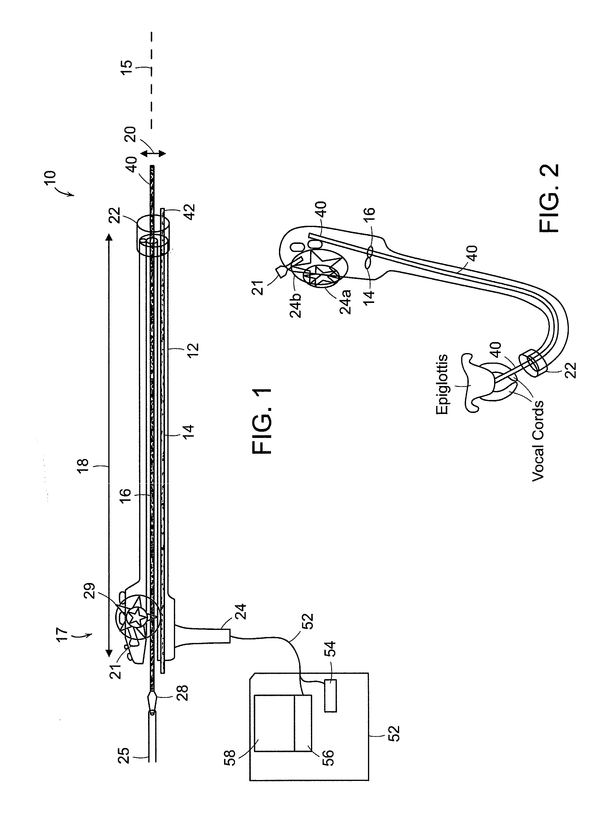
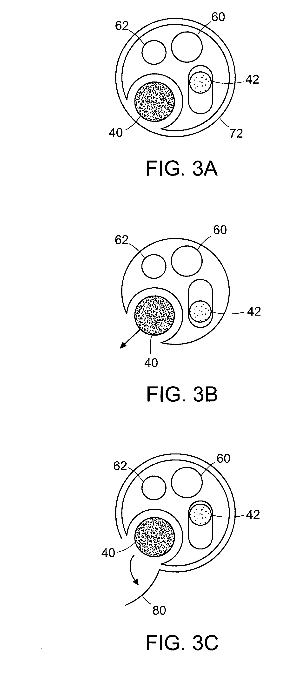
[0028]The present invention comprises an endotracheal endoscope 10 as illustrated in the schematic view of FIG. 1. The endoscope 10 has a flexible tubular body 12 extending along a central axis 15 with a first channel 16 in which a guide catheter 40 can be inserted. The endoscope 10 can have a width 20 in a range of 8-14 mm, preferably about 12 mm, and a length 18 in a range of 200-600 mm, preferably about 300-400 mm. The distal end of the endoscope can have a transparent distal window or clear cap 22 which can be retracted using a button at the proximal end control device 17. The control device can include a steering device or wheel 29, or two separate wheels, to control the direction of the distal end in two orthogonal directions. A second channel 14 can be used with a retraction catheter 42. The endoscope can be connected to a light source via a fiber optic cable 52 that extends from a light source 54 to the distal end of the endoscope where it is used to illuminate the field of ...


PUM


Abstract

The present invention relates to a guided endotracheal intubation endoscope. A guide catheter is used to position an endoscope and a tube for intubation of a patient. The endoscope can include a steering device for steering the endoscope in at least two directions, as well as suction, irrigation and retraction devices for maintaining a clear field of view.

Description

CROSS REFERENCE TO RELATED APPLICATIONS[0001]This application claims priority to U.S. Provisional Application 60 / 998,729 filed on Oct. 12, 2007, the entire contents of which is incorporated herein by reference.BACKGROUND OF THE INVENTION[0002]Tracheal intubation is a procedure involving the placement of a tube through the oral cavity, larynx, vocal cords and into the trachea. A laryngoscope is commonly used to view the glottis during placement of the endotracheal tube. The endotracheal tube often has a cuff that is inflated to form a seal with the interior walls of the trachea to prevent aspiration into the lungs.[0003]Endotracheal intubation in patients with a difficult airway is commonly performed using a fiberoptic or video bronchoscope or endoscope. The endoscope is passed through the vocal cords, and once the tracheal rings are identified, endotracheal position of the endoscope is presumed, and the endotracheal tube (ET) is passed blindly through the vocal cords into the trache...

Claims


Application Information

Patent Timeline
Patent Type & Authority Applications(United States)
IPC IPC(8): A61B1/267
CPCA61M16/0488A61B1/267A61M16/0486A61M16/0434A61B1/00131A61B1/0125A61B1/2673A61B1/2676A61B1/0052A61B1/0014A61B1/00101A61B1/018A61B1/00098
Inventor MATTHES, KAI
Owner BETH ISRAEL DEACONESS MEDICAL CENT INC
Learn More