Device for oral cavity examination
a technology for oral cavity and examination, which is applied in the field of devices for detecting abnormal epithelial tissue, can solve the problems of significantly higher relative hazards of death for patients who delay in obtaining a cancer consultation
- Summary
- Abstract
- Description
- Claims
- Application Information
AI Technical Summary
Benefits of technology
Problems solved by technology
Method used
Image
Examples
example 1
[0074]A routine visual examination of the oral cavity is made, noting the presence or absence of any lesions on the attached gingival, the buccal mucosa, the floor of the mouth, the hard and soft palate, and the dorsal, lateral, and ventral tongue. The presence or absence of any lesions noted by this routine examination are recorded. Additionally, the presence or absence of clusters of blood vessels (i.e., angiogenesis) which may indicate new growth such as cancer is noted.
example 2
[0075]After completing the routine examination of Example 1, the patient is then instructed to rinse the mouth with a 1% acetic acid solution for up to one minute and then expectorate.
[0076]Referring to FIG. 1, the device 110 of the present invention is activated by bending the sidewall 102 defining the chemical housing 114, thereby allowing the components therein to mix together.
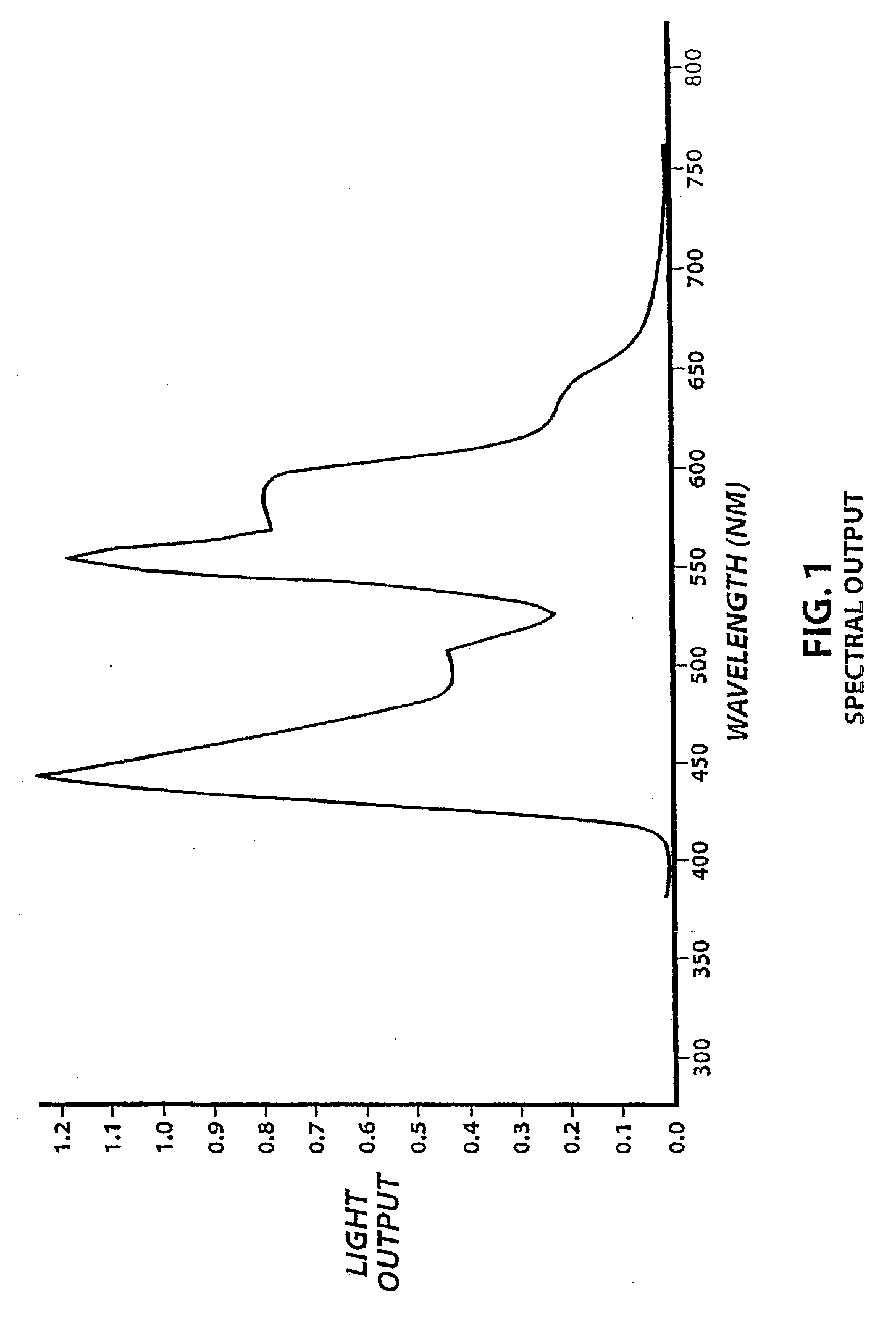
[0077]Preferably, and as indicated earlier, 9,10 diphenylanthracine (“DPHA”) is used as the light source, and the light provided has a spectral peak of about 430 nm. This spectral peak preferably produces a blue light. In a preferred embodiment, use of DPHA reduces the amount of mucosal glare and provides a softer light than the use of other chemiluminescent agents. In other embodiments, the chemiluminescent light source described in U.S. Pat. No. 5,329,938 to Lonky, the entire contents of which are herein incorporated by reference, may be used. The light source described in that patent is commercially avai...
example 3
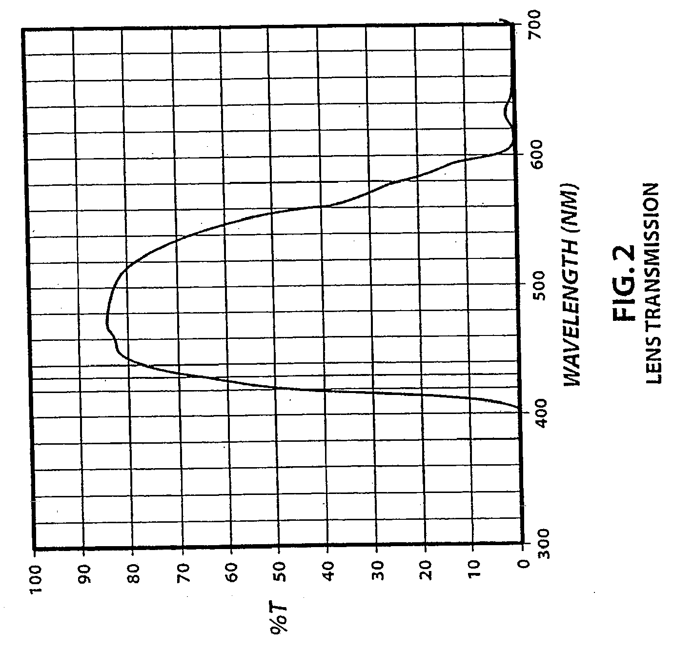
[0081]After completing the routine examination of Example 1, the patient is then instructed to rinse the mouth with a 1% acetic acid solution for up to a minute and then expectorate. Referring to FIGS. 1 and 2, the light source 100 of the device 110 of the present invention is then activated by bending the sidewall 102 defining the chemical housing 114.
[0082]The light source 100 emits light when activated. A portion of this light reflects off the reflective layer and travels back into the chemical housing. Preferably, at least a portion of this light is transmitted through the sidewall 102 defining the chemical housing 114. Additionally, a portion of the light emitted is incident light (as indicated earlier, collectively referred to herein as “emitted light”). Preferably, the light provided has a spectral peak at about 430 nm.
[0083]The emitted light is directed to the suspected pre-cancerous or cancerous region. The device is preferably manipulated so that little or none of the emit...
PUM
 Abstract
Description
Claims
Application Information
 - R&D Engineer
- R&D Manager
- IP Professional
- Industry Leading Data Capabilities
- Powerful AI technology
- Patent DNA Extraction
Browse by: Latest US Patents, China's latest patents, Technical Efficacy Thesaurus, Application Domain, Technology Topic, Popular Technical Reports.
© 2024 PatSnap. All rights reserved.Legal|Privacy policy|Modern Slavery Act Transparency Statement|Sitemap|About US| Contact US: help@patsnap.com


