
Method and system for bone suppression based on a single x-ray image

a single x-ray image and bone suppression technology, applied in the field of x-ray imaging, can solve the problems of difficult to detect lung nodules (i.e., potential lung cancer), difficult to detect nodules in chest radiographs, etc., and achieve the effect of suppressing bone structures, reducing the dimensionality of features, and reducing the set of features for each pixel

Inactive Publication Date: 2009-04-02
SIEMENS AG
View PDF8 Cites 25 Cited by
  • Summary
  • Abstract
  • Description
  • Claims
  • Application Information

AI Technical Summary

Benefits of technology

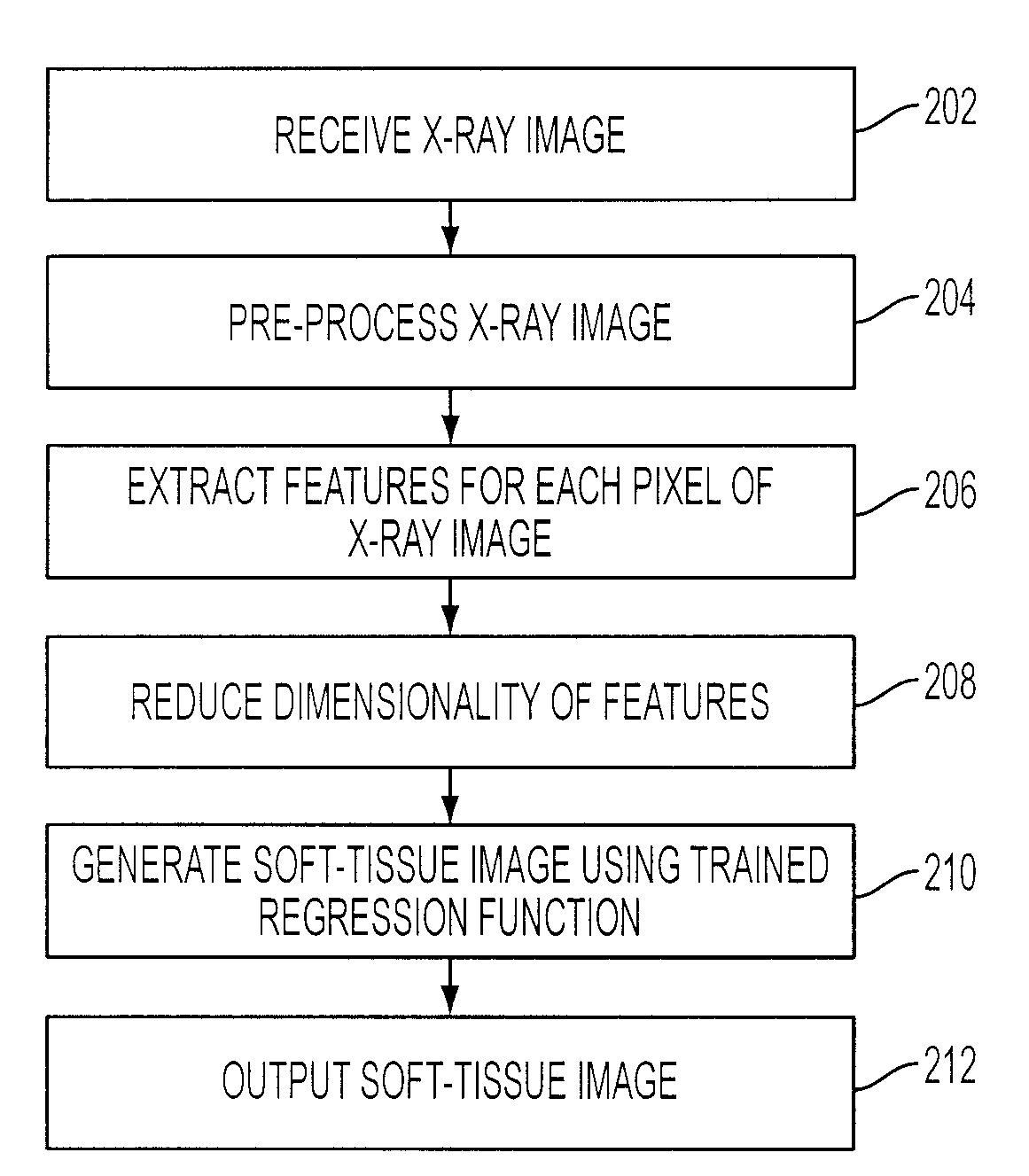
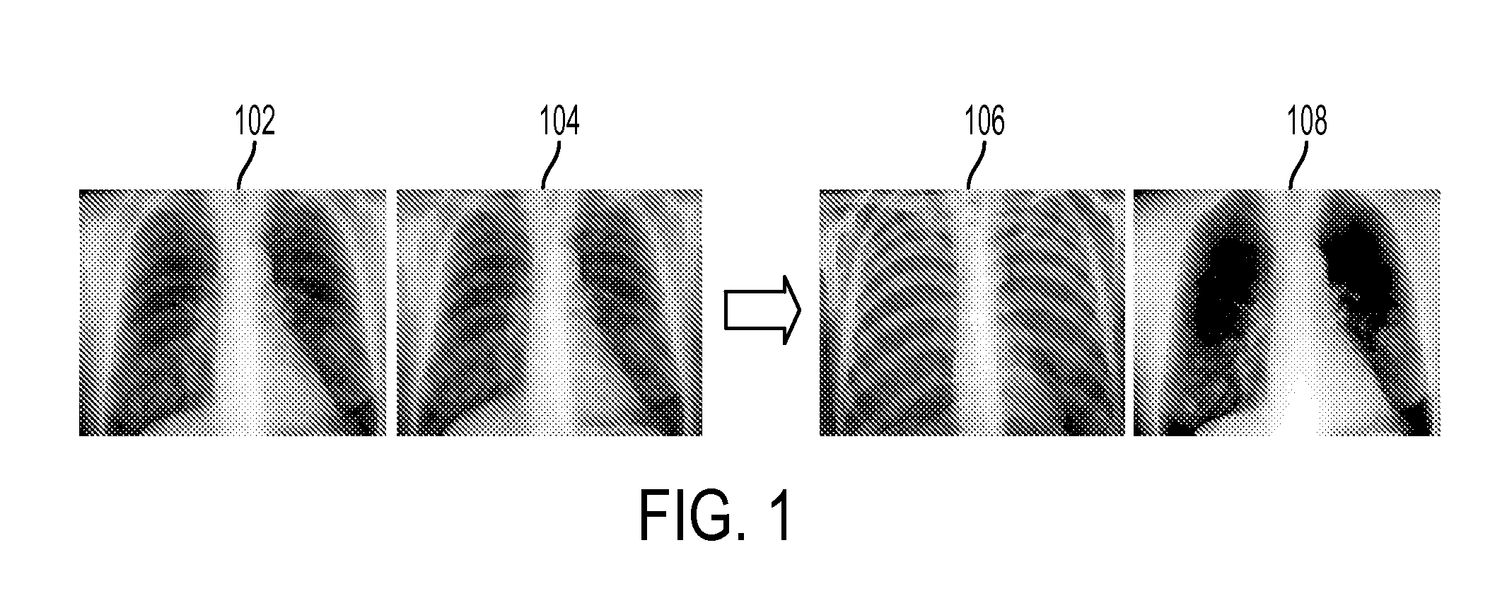
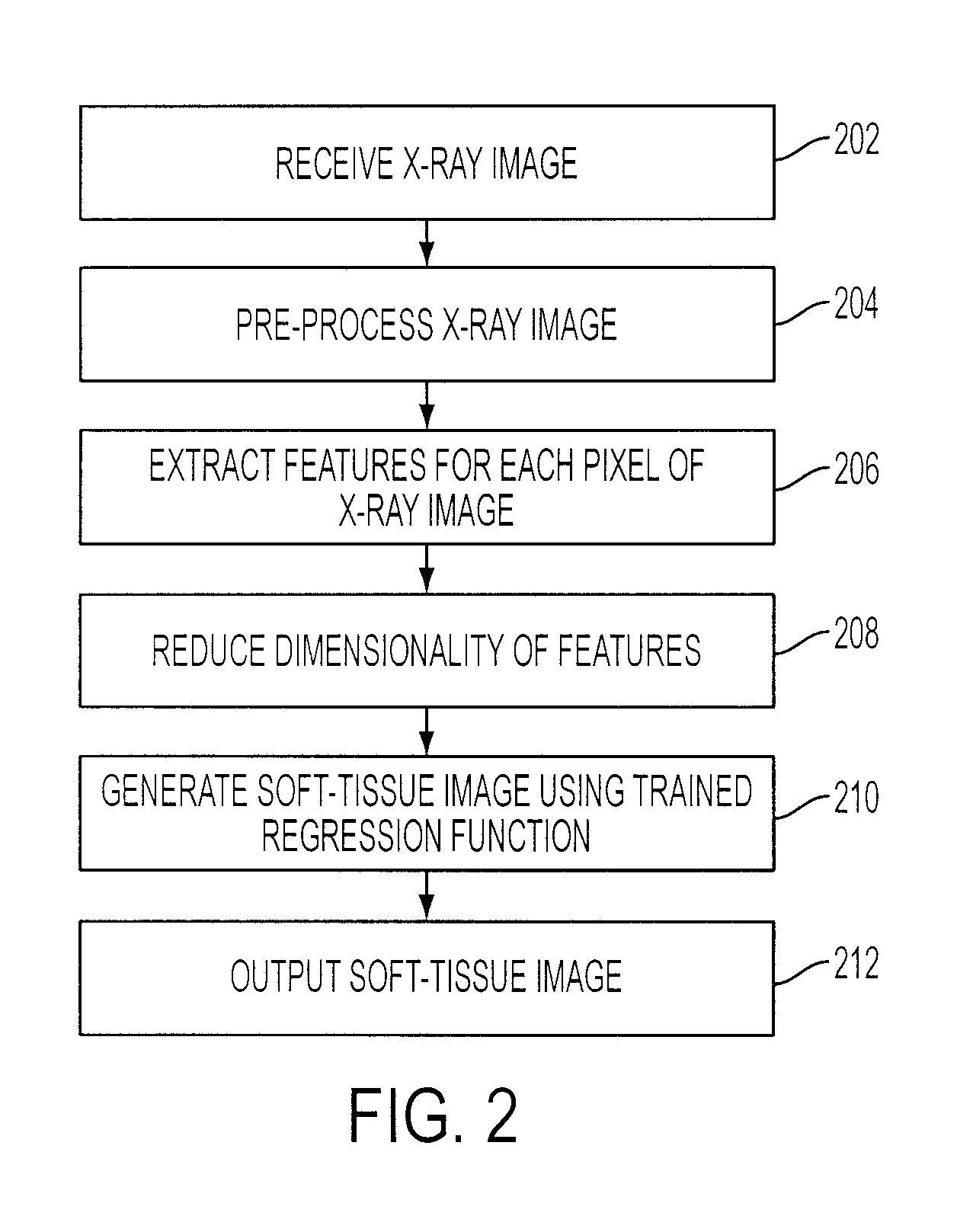
[0007]In one embodiment of the present invention, an x-ray image is received. A set of features is extracted for each pixel of the x-ray image. The extracted features may be wavelet features. A soft-tissue image is generated from the x-ray image using a trained regression function to determine an intensity value for the soft-tissue image corresponding to each pixel of the x-ray image based on the set of features extracted for each pixel of the x-ray image. The regression function can be trained using Bayesian Committee Machine (BCM) to approximate Gaussian process regression (GPR). The x-ray image may be normalized prior to extracting the features and the dimensionality of the set of features for each pixel may be reduced prior to generating the soft-tissue image.
[0008]In another embodiment of the present invention, multiple sets of training images, each set of training images having a training x-ray image and a corresponding training soft-tissue image, are received. A set of features, such as wavelet features, is extracted for each pixel in each of the training x-ray images. A regression function to suppress bone structures in x-ray images is trained based on the extracted wavelet features for the training x-ray images and the corresponding training soft-tissue images. The regression function may be trained using BCM to approximate GPR. The training x-ray images and the corresponding training soft-tissue images may be normalized prior to extracting the features, and the dimensionality of the features may be reduced prior to training the regression function.

Problems solved by technology

However, it is difficult to detect lung nodules (i.e., potential lung cancers) in a conventional chest radiograph (x-ray image) because lung nodules are often obscured in chest radiographs by overlying bones, such as ribs and clavicles.
Even with a computer-aided diagnostic (CAD) scheme for nodule detection, it can be challenging to detect nodules in chest radiographs when bones overlap with the nodules.

Method used


Image

  • Method and system for bone suppression based on a single x-ray image
  • Method and system for bone suppression based on a single x-ray image
  • Method and system for bone suppression based on a single x-ray image

Examples


Embodiment Construction

[0018]The present invention is directed to a method for suppressing bone structures in x-ray images. Embodiments of the present invention are described herein to give a visual understanding of the bone structure suppression method. A digital image is often composed of digital representations of one or more objects (or shapes). The digital representation of an object is often described herein in terms of identifying and manipulating the objects. Such manipulations are virtual manipulations accomplished in the memory or other circuitry / hardware of a computer system. Accordingly, is to be understood that embodiments of the present invention may be performed within a computer system using data stored within the computer system.

[0019]Embodiments of the present invention utilize a learning-based regression model for predictive bone suppression in an x-ray image. The regression model estimates a soft-tissue image from an input x-ray image (radiograph). The regression model is trained using...


PUM


Abstract

A method and system for suppressing bone structures based on a single x-ray image is disclosed. The bone structure suppressing method predicts a soft-tissue image without bone structures from an input x-ray image. A set of features is extracted for each pixel of the input x-ray image. A soft-tissue image is then generated from the input x-ray image using a trained regression function to determine an intensity value for the soft-tissue image corresponding to each pixel of the input x-ray image based on the set of features extracted for each pixel of the input x-ray image. The extracted features can be wavelet features and the regression function can be trained using Bayesian Committee Machine (BCM) to approximate Gaussian process regression (GPR).

Description

[0001]This application claims the benefit of U.S. Provisional Application No. 60 / 975,295, filed Sep. 26, 2007, the disclosure of which is herein incorporated by reference.BACKGROUND OF THE INVENTION[0002]The present invention relates to x-ray imaging, and more particularly, to suppressing bone structures based on a single x-ray image.[0003]Chest radiography (i.e., x-ray imaging) is a frequently used imaging technique for the diagnosis of chest diseases, such as lung cancer, pneumoconiosis, and pulmonary emphysema. However, it is difficult to detect lung nodules (i.e., potential lung cancers) in a conventional chest radiograph (x-ray image) because lung nodules are often obscured in chest radiographs by overlying bones, such as ribs and clavicles. Even with a computer-aided diagnostic (CAD) scheme for nodule detection, it can be challenging to detect nodules in chest radiographs when bones overlap with the nodules.[0004]A conventional solution to this problem is to use dual energy im...

Claims


Application Information

Patent Timeline
IPC IPC(8): G06K9/00
CPCG06T2207/10116G06T2207/20064G06T2207/30008G06T2207/30064G06T2207/20081G06T5/005G06T5/20G06T2207/20076G06T5/002G06T5/77G06T5/70G06T5/60
Inventor SLABAUGH, GREGORY G.FANG, TONGMOON, KOOKSANGCHEN, YUNQIANG
Owner SIEMENS AG
Learn More