
Apparatus and methods for internal surgical procedures

a surgical device and internal surgical technology, applied in the field of internal surgical devices and methods, can solve the problems of difficult removal of superficial tumors, internal hemorrhoids, polyps, etc., and the difficulty of the practitioner resecting large sessile polyps and other lesions without injuring

Active Publication Date: 2009-03-12
MAYO FOUND FOR MEDICAL EDUCATION & RES
View PDF32 Cites 228 Cited by
  • Summary
  • Abstract
  • Description
  • Claims
  • Application Information

AI Technical Summary

Benefits of technology

[0004]In the absence of a submucosal liquid cushion, it may be difficult for the practitioner to resect large sessile polyps and other lesions without injuring the underlying muscularis propria. By raising the lesion with a submucosal liquid cushion, the lesion may be isolated from the surrounding tissue and the underlying muscularis propria may be better protected from injury during resection of the lesion.
[0005]After the submucosal liquid cushion is formed, resection may be performed using a snare looped around the base of the submucosal liquid cushion. Examples of snare resection devices are described in, e.g., U.S. Pat. Nos. 5,542,948 (Weaver et al.) and 6,210,416 B1 (Chu et al.). Resection using the snare devices is typically augmented by electrosurgical energy to enhance cutting and, in some instances, to reduce bleeding.SUMMARY OF THE INVENTION
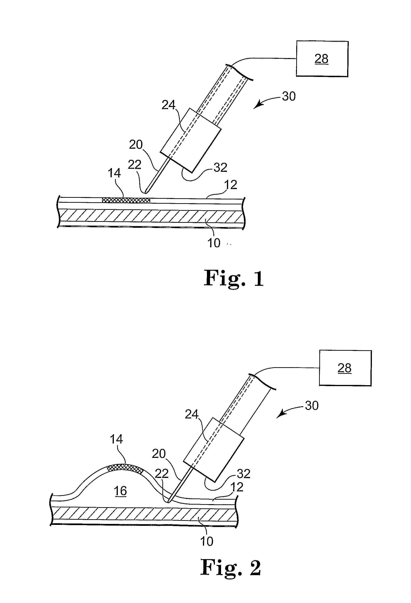
[0009]The apparatus and methods for creating blebs may preferably involve the delivery of gas submucosally to separate tissue and create a gas-filled submucosal space. The gas may preferably be delivered at a pressure greater than atmospheric pressure. Using pressurized gas to create gas-filled blebs may provide a number of advantages over blebs formed using liquids. It has been observed that blebs created with pressurized gas may be higher, i.e., the gas-filled submucosal space may potentially be higher when measure normal to the plane of the underlying tissue. It has also been observed that tissue separation within the space of a gas-filled bleb may potentially be more pronounced than within a liquid-filled bleb.
[0010]Another potential advantage of a gas-filled bleb is that use of an electrosurgical cutting apparatus designed for “inside-out” resection may be more effective when used in connection with a gas-filled bleb. As described herein, some resection devices of the present invention may be effective when used to perform “inside-out” resection of tissue. Unlike conventional mucosal tissue resection in which a cutting instrument is used to cut tissue from the outside of a bleb towards the submucosal space within the bleb, methods of the present invention may preferably involve piercing the tissue of a bleb with a resection device and advancing at least a portion of the resection into the submucosal space. After the resection device (or a portion thereof) is located within the submucosal space, the tissue raised within the bleb can be cut from the inside of the submucosal space outwardly. By directing a cutting action outwardly from within the bleb, cutting and / or perforation of the tissue underlying the bleb may be much less likely to occur.
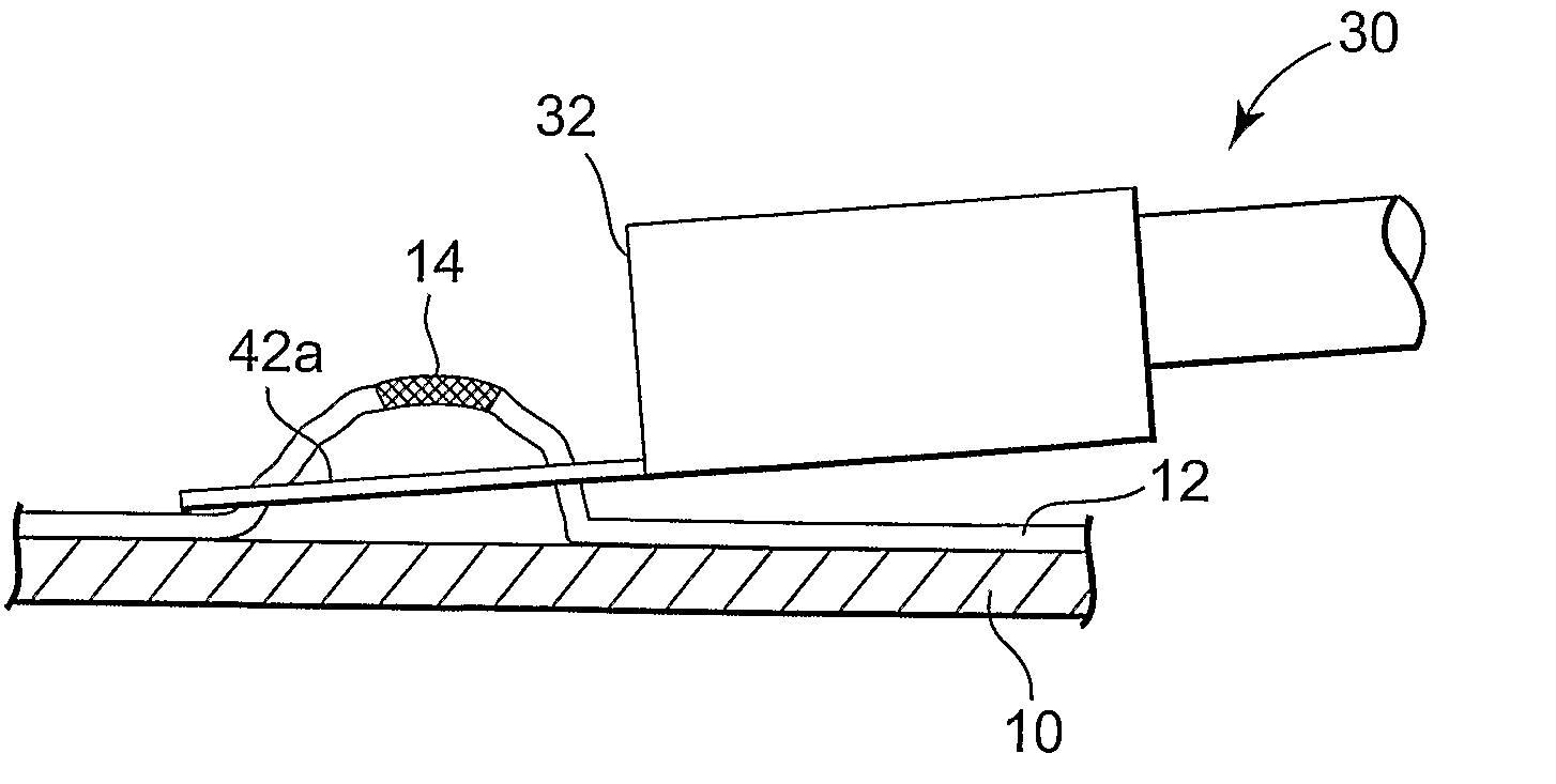
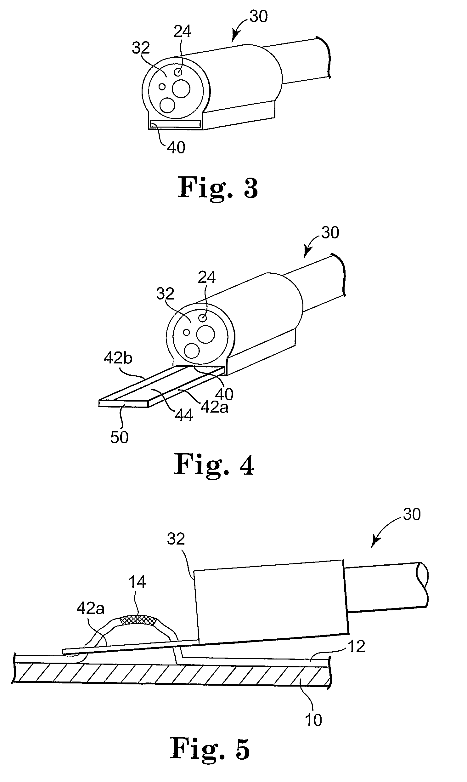
[0020]The use of a resection frame as discussed in connection with the present invention may be advantageous in connection with a gas delivery system for forming blebs. For example, the resection frame may be extended and placed around the tissue to be resected. When in position, the resection frame can be used to apply pressure about the perimeter of the location at which a bleb is to be formed. The pressure provided by the resection frame may serve to restrict the size of the bleb by reducing the likelihood that the gas will separate tissue under the frame.

Problems solved by technology

A variety of lesions such as superficial tumors, polyps, internal hemorrhoids, etc. may be difficult to remove from internal body locations because they have a relatively low profile with respect to surrounding tissue.
In the absence of a submucosal liquid cushion, it may be difficult for the practitioner to resect large sessile polyps and other lesions without injuring the underlying muscularis propria.

Method used


Image

  • Apparatus and methods for internal surgical procedures
  • Apparatus and methods for internal surgical procedures
  • Apparatus and methods for internal surgical procedures

Examples


Embodiment Construction

[0085]In the following detailed description of some exemplary embodiments of the invention, reference is made to the accompanying figures which form a part hereof, and in which are shown, by way of illustration, specific embodiments in which the invention may be practiced. It is to be understood that other embodiments may be utilized and structural changes may be made without departing from the scope of the present invention.

[0086]The present invention may, in various embodiments, include three basic components, an injection apparatus capable of creating a submucosal fluid cushion, a resection apparatus capable of resecting tissue raised above the submucosal fluid cushion, and a stapling apparatus capable of stapling tissue as a part of the tissue removal process. It may be preferred that all three components, i.e., the injection apparatus, resection apparatus, and stapling apparatus be combined in the same instrument as depicted in many of the figures described below. It should, ho...


PUM

PropertyMeasurementUnit
angleaaaaaaaaaa
angleaaaaaaaaaa
pressureaaaaaaaaaa

Abstract

Apparatus and methods for internal surgical procedures are disclosed. The apparatus and methods may involve supporting internal body locations, creating submucosal separations (blebs), and / or for resecting mucosal tissue separated from underlying tissue by a bleb.

Description

RELATED APPLICATION[0001]The present application claims the benefit under 35 U.S.C. §119(e) of U.S. Provisional Patent Application No. 60 / 679,760 filed May 11, 2005 and titled APPARATUS AND METHODS FOR INTERNAL SURGICAL PROCEDURES, which is hereby incorporated by reference in its entirety.[0002]The present invention relates to internal surgical devices and methods. More particularly, the present invention relates to devices that may beneficially be used in conjunction with, e.g., mucosectomy procedures.[0003]A variety of lesions such as superficial tumors, polyps, internal hemorrhoids, etc. may be difficult to remove from internal body locations because they have a relatively low profile with respect to surrounding tissue. The injection of liquids into the submucosa may be performed to facilitate surgical procedures such as, e.g., endoscopic mucosal resection (EMR), polypectomies in the gastrointestinal tract. The injected liquid may advantageously form a submucosal cushion beneath ...

Claims


Application Information

Patent Timeline
Patent Type & Authority Applications(United States)
IPC IPC(8): A61B17/32A61B17/94A61B17/072A61M29/00A61M5/158A61B18/14
CPCA61B17/221A61B2017/320044A61B2017/00269A61B2017/00296A61B2017/22082A61B2017/2212A61B2017/2929A61B2018/1495A61B17/0218A61B17/0644A61B17/072A61B17/320016A61B18/1445A61B2017/00318A61B2017/00323A61B2017/00867A61B2017/00986A61B2017/07214A61B2017/07271A61B2018/141A61B2019/481A61B2019/4878A61B1/018A61B18/1492A61B17/32056A61B2090/08021A61B2090/0815
Inventor DE LA MORA LEVY, JOSE G.GOSTOUT, CHRISTOPHER J.
Owner MAYO FOUND FOR MEDICAL EDUCATION & RES
Learn More