
Method and system for displaying tomosynthesis images

a tomosynthesis and image technology, applied in the field of tomosynthesis, can solve the problem that the practitioner cannot acquire all of the information, and achieve the effect of facilitating the acquisition of information

Active Publication Date: 2009-02-05
GENERAL ELECTRIC CO
View PDF4 Cites 90 Cited by
  • Summary
  • Abstract
  • Description
  • Claims
  • Application Information

AI Technical Summary

Benefits of technology

"The invention proposes a method for displaying images obtained by tomosynthesis, which involves taking multiple X-ray images at different angles and combining them to create a three-dimensional image. The method allows for the selection and display of specific volumes of interest within the image data. The resulting images can be used for diagnosis and treatment purposes. The invention also includes a system for displaying and managing the tomosynthesis images, as well as a computer program for implementing the method. The technical effects of the invention include improved image quality, improved diagnostic accuracy, and improved treatment precision."

Problems solved by technology

Consequently, visualizing one given slice of a radiological sign does not allow the practitioner to acquire all of the information relative to this sign.

Method used


Image

  • Method and system for displaying tomosynthesis images
  • Method and system for displaying tomosynthesis images
  • Method and system for displaying tomosynthesis images

Examples


Embodiment Construction

[0055]In the case of DBT imaging, images of the patient's breast are taken at different angles so as to be able to reconstruct a three-dimensional representation of the breast. Once reconstructed, this three-dimensional representation makes it possible to observe and localize the internal structures of the breast. It is typically made up of a set of slices parallel to the detector, which, superimposed, represent the volume of the compressed breast. In these slices, the practitioner can detect anomalies such as opacities, microcalcification foci, which can be benign or malignant lesions.

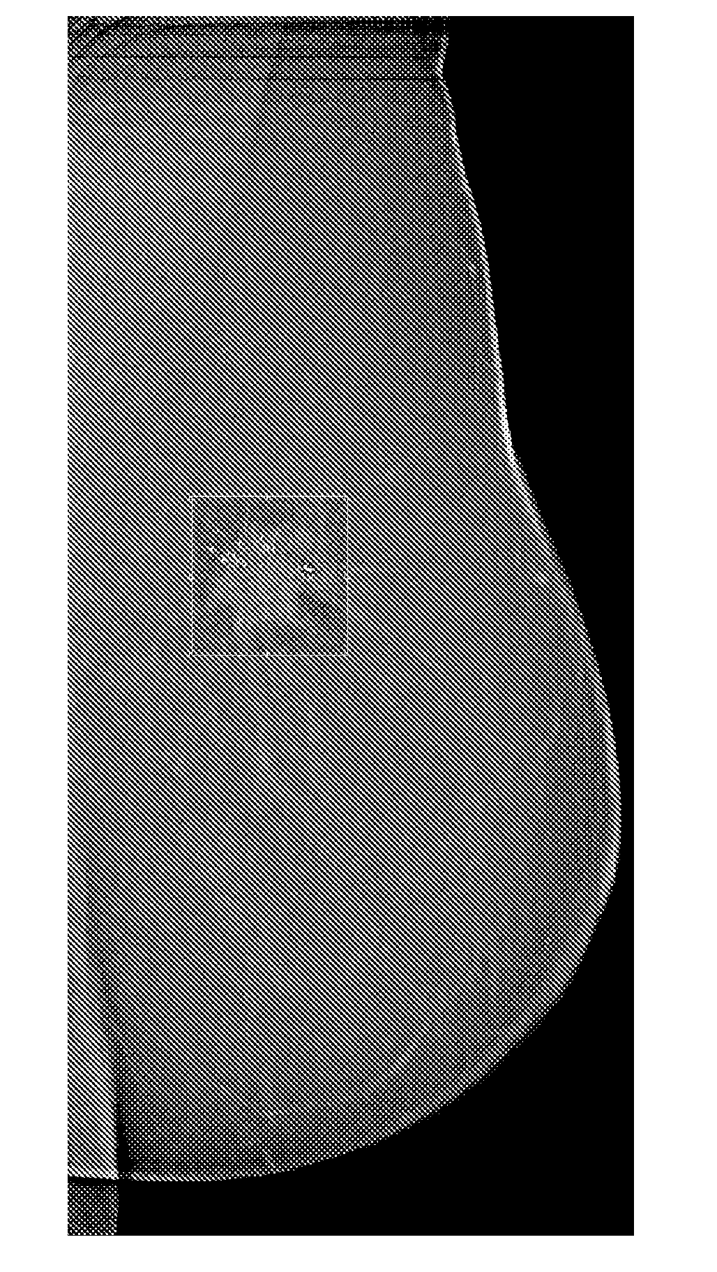
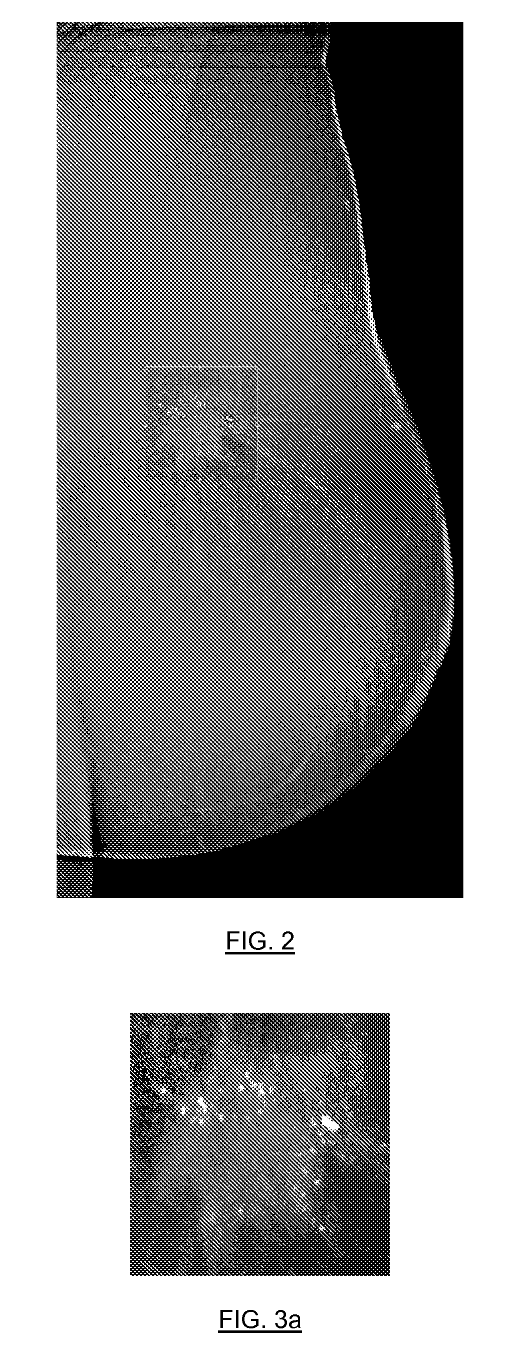
[0056]As illustrated in FIG. 2, it is proposed to the practitioner to display, in a window, on a two-dimensional image which can be an image of a slice or a slab, thumbnail images corresponding to a region of particular interest including, for example, a lesion which the practitioner will have been able to detect and wishes to analyze more specifically. In this window, a particular local display such ...


PUM


Abstract

An embodiment of a method for displaying a volume obtained by tomosynthesis includes displaying a two-dimensional image. It further includes receiving user input that defines on the displayed image at least one volume of interest associated with a two-dimensional region of interest located in a plane of the image. The method further includes displaying in the region of interest, according to a practitioner's wishes: (a) images of slices of the volume of interest; (b) three-dimensional images of the volume of interest; and / or (c) slabs obtained from the volume of interest.

Description

CROSS-REFERENCE TO RELATED APPLICATIONS[0001]This application claims the benefit of priority, under 35 U.S.C. 119(a)-(d), (f) to prior-filed French patent application serial number 0756896, filed on 2 Aug. 2007, the contents of which are herein incorporated by reference in their entirety.FIELD OF THE INVENTION[0002]The field of the invention relates to tomosynthesis generally, and in particular to a method and a system for displaying volumes obtained by tomosynthesis.[0003]It is applicable especially, but not exhaustively, to a 3D radiography imaging technique of the breast, called DBT (Digital Breast Tomosynthesis), or other organs in traditional radiology.BACKGROUND TO THE INVENTION[0004]Tomosynthesis imagery is a three-dimensional imaging technique using limited-angle tomography. This technique makes it possible to reconstruct a three-dimensional (3D) volume in slices from a series of bi-dimensional (2D) projection images acquired using different angles of the X-ray tube.[0005]In...

Claims


Application Information

Patent Timeline
Patent Type & Authority Applications(United States)
IPC IPC(8): H05G1/64
CPCG06T19/00G06T2219/028G06T2210/41
Inventor BERNARD, SYLVAINCORGNE, BRUNO LEPETER, FRANCOIS
Owner GENERAL ELECTRIC CO
Learn More