
Method for determining an optimal output of an ablation catheter for a myocardial ablation in a patient and associated medical apparatus

Active Publication Date: 2008-12-25
SIEMENS HEALTHCARE GMBH
View PDF10 Cites 8 Cited by
  • Summary
  • Abstract
  • Description
  • Claims
  • Application Information

AI Technical Summary

Benefits of technology

[0024]It is thus possible, with knowledge of the thickness of the myocardium, in some instances at the current position of the ablation catheter, to determine the output of the latter optimally point by point and where necessary to set it accordingly. This protects healthy tissue, since it is ablated with optimal and not excessive output. The success rate for an ablation intervention is increased, since the likelihood of not reaching pathological neuron pathways in the epicardium is minimized.
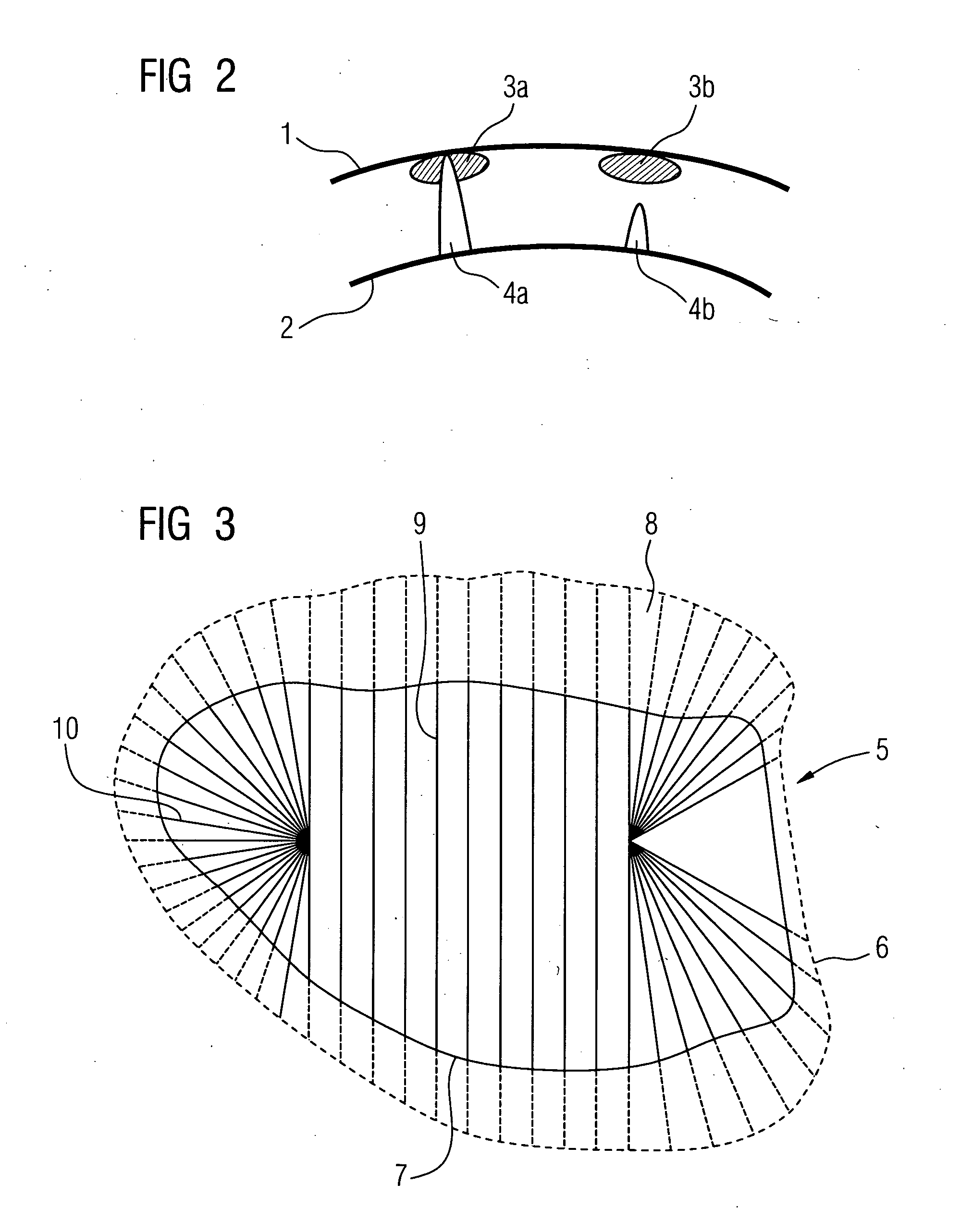
[0037]For fast acquisition of the depth, in particular from a qualitative point of view, a color coding is recommended such that regions with a thin myocardium are colored red for example, while regions with a greater myocardium thickness are shown as blue. Intermediate regions can be marked accordingly with transition colors. This means that the very significant variations in myocardium thickness, which can be between 0.4 cm and 2 cm in individual instances in one region, can be quickly acquired. Information about the different myocardium thicknesses is therefore displayed directly to an operator or the ablation catheter system. This information is extremely important, since successful ablation can only be achieved even with normal fluctuations in thickness in the region of around 0.8 cm to 1.4 cm, if catheter output is actually tailored precisely to these thickness differences.
[0039]The location-dependent thickness of the myocardium in the ablation region and / or in some instances a representation of the endocardial surface can be registered with at least one current position information item of the ablation region, in particular by the computation apparatus or in some instances a computation apparatus of the ablation catheter system and / or by means of current image recordings of the ablation region. Of course only one integrated computation apparatus may also be provided. Registration of the surface model with current position information for the ventricle allows assignment of the depth information to the region currently of importance for the treatment or the region acquired by this. The thickness information, which was obtained for example by means of a computed tomography scan obtained one or two days before the actual ablation, can thus be compared with the current position of the ventricle. This ensures that the locally correct thickness information is used for the ablation.
[0042]The image recording apparatus used to create the at least one at least three-dimensional image recording can be a computed tomography apparatus and / or a magnetic resonance apparatus and / or a rotation angiography apparatus and / or an ultrasound apparatus. Other imaging techniques not mentioned here can also be used, which similarly allow the creation of three or multi-dimensional image recordings, for example from films of three-dimensional images. It is important that one at least three-dimensional recording of the heart or left myocardium is possible. It is also possible for the three-dimensional image recordings or multi-dimensional image recordings to be combined or merged from individual recordings using different imaging techniques, so that further information from another technique can be included in magnetic resonance images for example, to improve image quality.

Problems solved by technology

Abnormalities of pathological origin here cause defective neuron pathways in the ventricle with the result that the heart beats in an uncontrolled manner (too frequently).
This is associated with a reduction in the time available to fill the ventricle, so cardiac performance is greatly reduced.
Ventricular fibrillation or flutter gives rise to acute complaints such as chest pain and reduced capability, etc.
There is also an increased risk of thrombosis, in turn significantly increasing the likelihood of stroke.
One problem with such ablation methods is that the myocardium or cardiac muscle is not of uniform thickness but its thickness varies locally and with each patient.
If the output is too low, it may be that pathological pathways in the epicardium, in other words in the outer regions of the myocardium, are not reached and the ablation is therefore unsuccessful.
If catheter output is too high on the other hand, unnecessary scarring of the myocardial tissue may result and in some instances the pericardium or heart sac may even be damaged.
For these reasons ablation of the left cardiac muscle or myocardium is currently rarely undertaken, as there is a high probability that pathological pathways in the epicardium for example will not be reached.
It is not possible to identify this thickness in the fluoroscopy image.
The physician is therefore only able to use mean values to set the ablation output, with the result that the problems mentioned above arise.
It is not possible to ensure optimal setting of output with such an estimation of the thickness of the myocardium and appropriate output.

Method used


Image

  • Method for determining an optimal output of an ablation catheter for a myocardial ablation in a patient and associated medical apparatus
  • Method for determining an optimal output of an ablation catheter for a myocardial ablation in a patient and associated medical apparatus
  • Method for determining an optimal output of an ablation catheter for a myocardial ablation in a patient and associated medical apparatus

Examples


Embodiment Construction

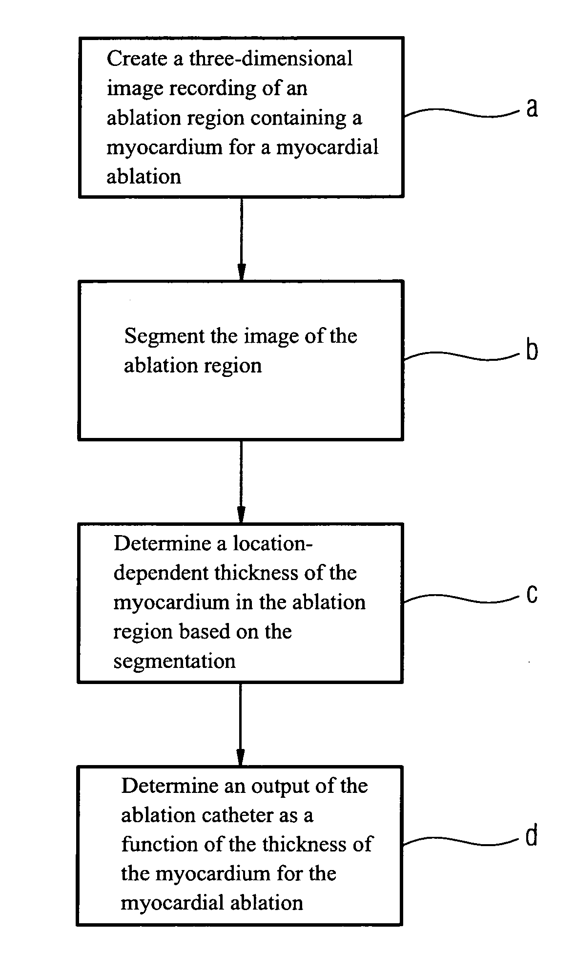
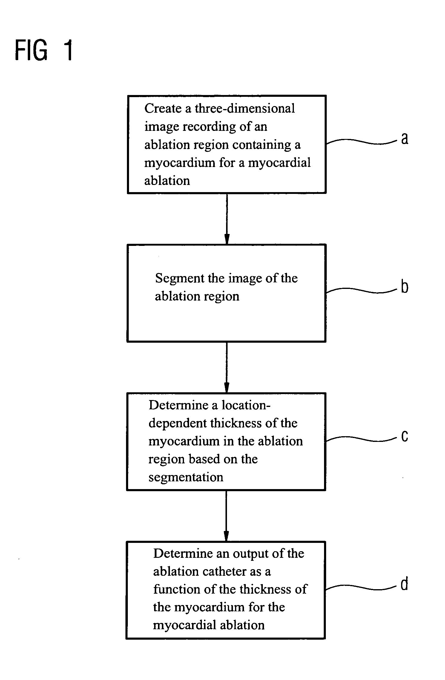
[0053]FIG. 1 shows an outline of the sequence of an inventive method.

[0054]According to step a at least one at least three-dimensional image recording is first created of at least one ablation region provided for the myocardial ablation, in other words for example of a left ventricle of a patient, with the aid of an image recording apparatus or a number of image recording apparatuses, such as a computed tomograph or a magnetic resonance apparatus. Images can likewise be created as part of rotation angiography or using ultrasound.

[0055]Then according to step b an at least partial segmentation of the image recordings or of the recorded ablation region is carried out. Segmentation, in other words the division of the content, allows segmentation information to be obtained, with segmentation being carried out by a computation apparatus, in some instances with appropriate software means.

[0056]In step c the segmentation information is used to calculate the location-dependent thickness of t...


PUM


Abstract

The invention relates to a method for determining an optimal output of an ablation catheter for a myocardial ablation in a patient with the following steps: creation of at least one at least three-dimensional image recording of an ablation region provided for the myocardial ablation using at least one image recording apparatus; at least partial segmentation of the recorded ablation region to obtain segmentation information using a computation apparatus; at least partial determination from the segmentation information of the location-dependent thickness of the myocardium in the ablation region by the computation apparatus; and determination of the optimal output of the ablation catheter, in particular by the computation apparatus or a separate computation apparatus of an ablation catheter system, as a function of the determined myocardium thickness.

Description

CROSS REFERENCE TO RELATED APPLICATIONS[0001]This application claims priority of German application No. 10 2007 028 115.5 filed Jun. 19, 2007, which is incorporated by reference herein in its entirety.FIELD OF THE INVENTION[0002]The invention relates to a method for determining an optimal output of an ablation catheter for a myocardial ablation in a patient and associated medical apparatus.BACKGROUND OF THE INVENTION[0003]According to current studies the number of cardiological diseases is constantly increasing. One typical pathological finding here is so-called ventricular fibrillation or flutter. Abnormalities of pathological origin here cause defective neuron pathways in the ventricle with the result that the heart beats in an uncontrolled manner (too frequently). This is associated with a reduction in the time available to fill the ventricle, so cardiac performance is greatly reduced.[0004]Ventricular fibrillation or flutter gives rise to acute complaints such as chest pain and ...

Claims


Application Information

Patent Timeline
IPC IPC(8): G06K9/00
CPCA61B18/1492A61B90/36A61B34/10
Inventor LAUTENSCHLAGER, STEFAN
Owner SIEMENS HEALTHCARE GMBH
Learn More