Thioflavin t method for detection of amyloid polypeptide fibril aggregation
a technology of amyloid polypeptide and thioflavin, which is applied in the field ofthio, can solve the problems of difficult interpretation, hampered applicability, and quantitative relationship
- Summary
- Abstract
- Description
- Claims
- Application Information
AI Technical Summary
Benefits of technology
Problems solved by technology
Method used
Image
Examples
example 1
[0025]This Example describes an illustrative method for performing the steps of the method of the invention.
[0026](1) Prepare a stock ThT solution (1 mg / ml; 3.14 mM) in distilled, deionized water, aliquot into small samples and store long term at −20° C. in the dark. Thaw frozen samples only once and discard the remainder. Add 1.6 μl of ThT solution to 1.0 ml of 50 mM Tris buffer, pH 7.4 to yield a final 5.0 μM ThT solution.
[0027](2) β-amyloid peptide (1-40) can be obtained from Biosource (Camarillo, Calif.), CLN from ReGen Therapeutics Plc (London, UK) and Thioflavin T from Sigma (St. Louis, Mo.). Prepare a stock solution of β-amyloid (by dissolving in 10 mM NaOH (0.5 mg / ml) and stored at −70° C. until used. Aβ in base is stable for at least 3 weeks frozen. Add 5.0 μl (2.5 μg) of β-amyloid stock solution to 1.0 ml of ThT / Tris buffer solution. It is preferable to avoid any contact with metal when preparing a amyloid solution since metal can oxidize the peptide.
[0028](3) Measure the ...
example 2
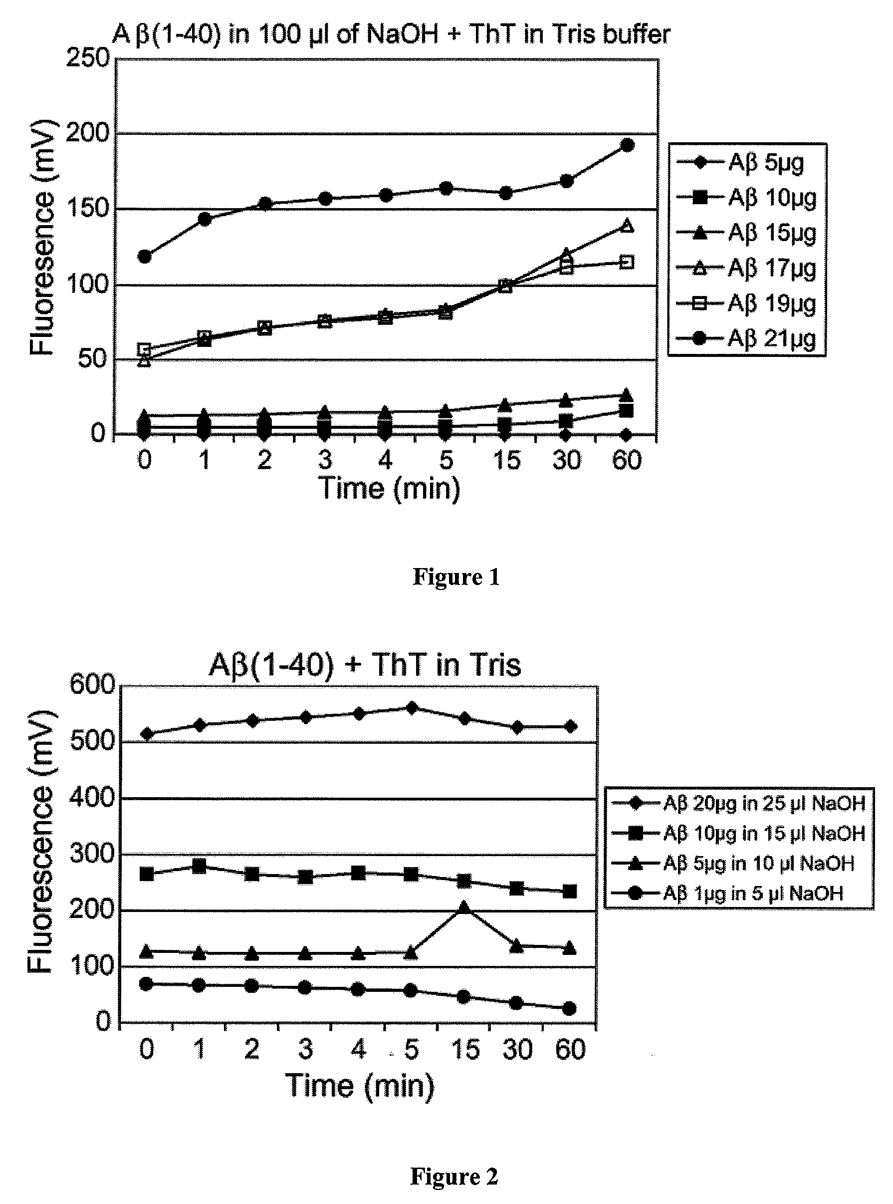
[0031]This example describes the relationship of ThT fluorescence to Aβ amount when the stoichiometries of Aβ to base were outside of the present invention. Increasing amounts of Aβ40 was added to 100 μl of 10 mM NaOH, which was then transferred to 1.0 ml of Tris buffer at pH 7.4 and containing 5 μM ThT. As can be seen in FIG. 1, the fluorescence is not proportional to the amount of Aβ40 peptide. Little fluorescence is observed at 5 and 10 μg of peptide and a significant increase in fluorescence takes place at 15 μg, but is not distinguishable from 17 μg of Aβ40. The stoichiometries for Aβ to base ranged from 1:20 to about 1:5.
example 3
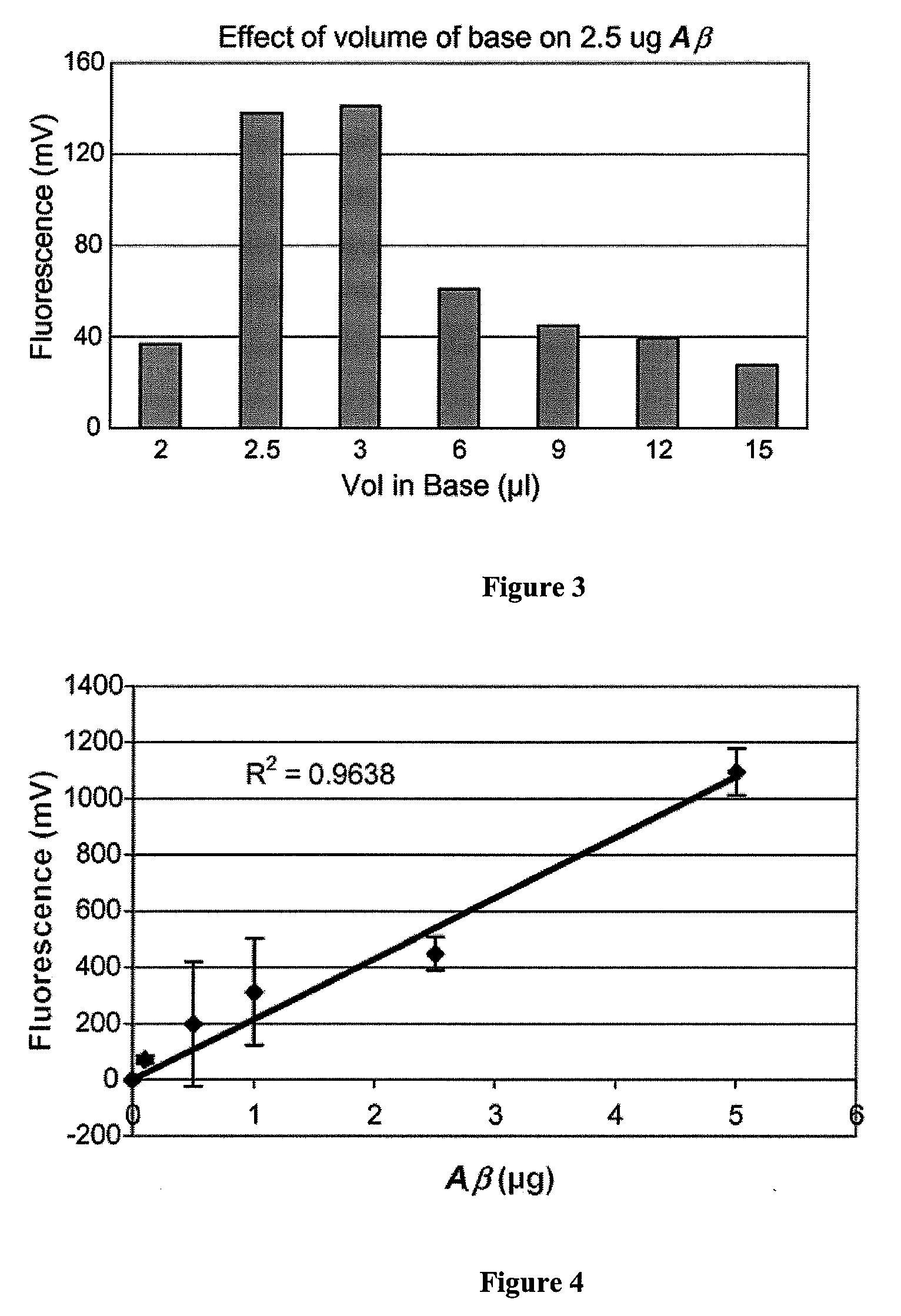
[0032]In order to test whether aggregation failed to take place due to the relatively large volume of base used to solubilize the smaller quantities of Aβ, a relatively constant ratio of peptide to the volume of base was maintained. As shown in FIG. 2, reducing the volume of base dramatically increases the resultant fluorescence at all concentration of peptide used. At the 5 μg / 100 μl NaOH used in FIG. 1, fluorescence was negligible. Upon reducing the volume of base to 10 μl, the same 5 μg of peptide demonstrated a fluorescence of 300 mV, which is higher than that observed for 21 μg (˜120 mV) of Aβ dissolved in 100 μl of base in FIG. 1. Thus, by reducing the volume of base, an appreciable level of fluorescence was observed down to the 1 μg of Aβ. Furthermore, fluorescence remained constant after the first few seconds of oligomerization as opposed to FIG. 1, whereby the fluorescence intensity continued to increase over the 60 min using the larger volume of base.
PUM
| Property | Measurement | Unit |
|---|---|---|
| time | aaaaa | aaaaa |
| pH | aaaaa | aaaaa |
| emission wavelength | aaaaa | aaaaa |
Abstract
Description
Claims
Application Information
 - R&D
- Intellectual Property
- Life Sciences
- Materials
- Tech Scout
- Unparalleled Data Quality
- Higher Quality Content
- 60% Fewer Hallucinations
Browse by: Latest US Patents, China's latest patents, Technical Efficacy Thesaurus, Application Domain, Technology Topic, Popular Technical Reports.
© 2025 PatSnap. All rights reserved.Legal|Privacy policy|Modern Slavery Act Transparency Statement|Sitemap|About US| Contact US: help@patsnap.com


