PTEN/Akt methods and compositions relating to BMP
a technology of pten/akt and composition, applied in the field of molecular biology, can solve the problems of insufficient vivo evidence, failure of most current therapies to completely cure cancer, and lack of direct linkage of mutated stem cells with initiation of neoplasms in the natural setting, and achieve the effect of increasing the number of crypts
- Summary
- Abstract
- Description
- Claims
- Application Information
AI Technical Summary
Benefits of technology
Problems solved by technology
Method used
Image
Examples
example 1
Induced Inactivation Of PTEN Leads To Development of Intestinal Polyposis
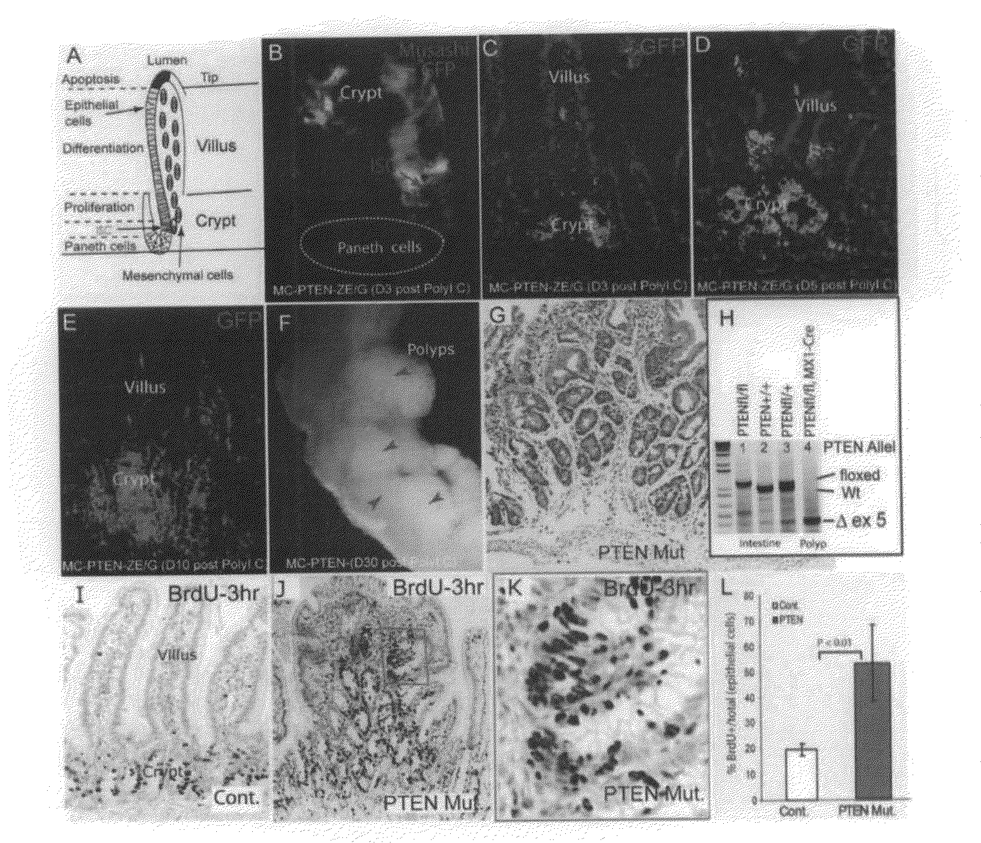
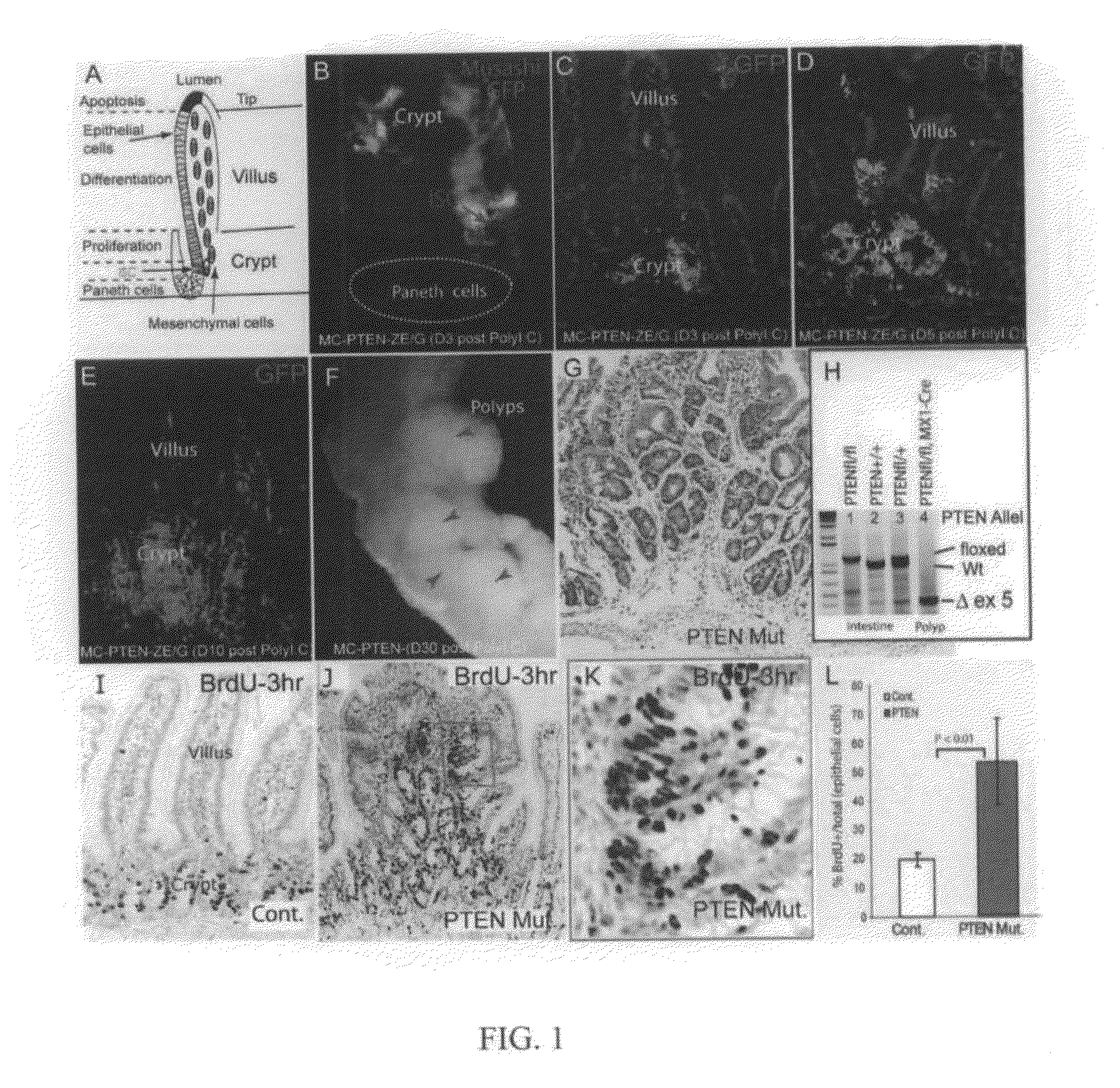
[0149]To determine the time-course of PTEN loss in the conditional deletion system, Mx1-Cre: Ptenfl / fl mice carrying a Z / EG reporter were generated, so that lineages from which Cre activity-deleted PTEN would be marked by the expression of green fluorescent protein (GFP). Gene deletion was induced at or before weaning with pIpC (see Methods above). GFP+ cells were first detected three days after completion of pIpC treatment (day 3). Co-staining with the stem cell marker Musashi1 (Musashi) confirmed that recombination had occurred in ICS (FIG. 1B). Initially, GFP+ epithelial cells were primarily clustered in the crypt bottom or located at the transition zone between crypts and villi (FIG. 1B, 1C). The GFP+ domains expanded to include groups of crypts on day 5 (FIG. 1D), and finally were seen to encompass some entire crypts and villi on day 10 (FIG. 1E). A limited number of stromal cells were also shown to be GFP...
example 2
PTEN Controls Cell Cycle in Intestinal Crypts
[0151]PTEN is known to inhibit cell proliferation through suppression of PI3K / Akt activity. Therefore, the proliferation state of cells in the intestinal polyp regions of the PTEN mutants was assessed by labeling actively cycling cells with a 3-hour pulse of BrdU. In normal / wild type (wt) crypts, BrdU+ cells were limited to a narrow proliferation zone, the TAC, (FIG. 1I). In PTEN mutant polyps, however, the BrdU+ cells were spread widely within the multiple crypt-like structures (FIG. 1J), the proliferating tissue appeared hyperplastic compared to the linear arrangement of proliferating cells in normal crypts (FIG. 1K), and the proliferative index of the epithelial cells (BrdU+ / (BrdU++BrdU−)) was substantially increased (2.7 fold, P<0.01) compared to wt control (FIG. 1G).
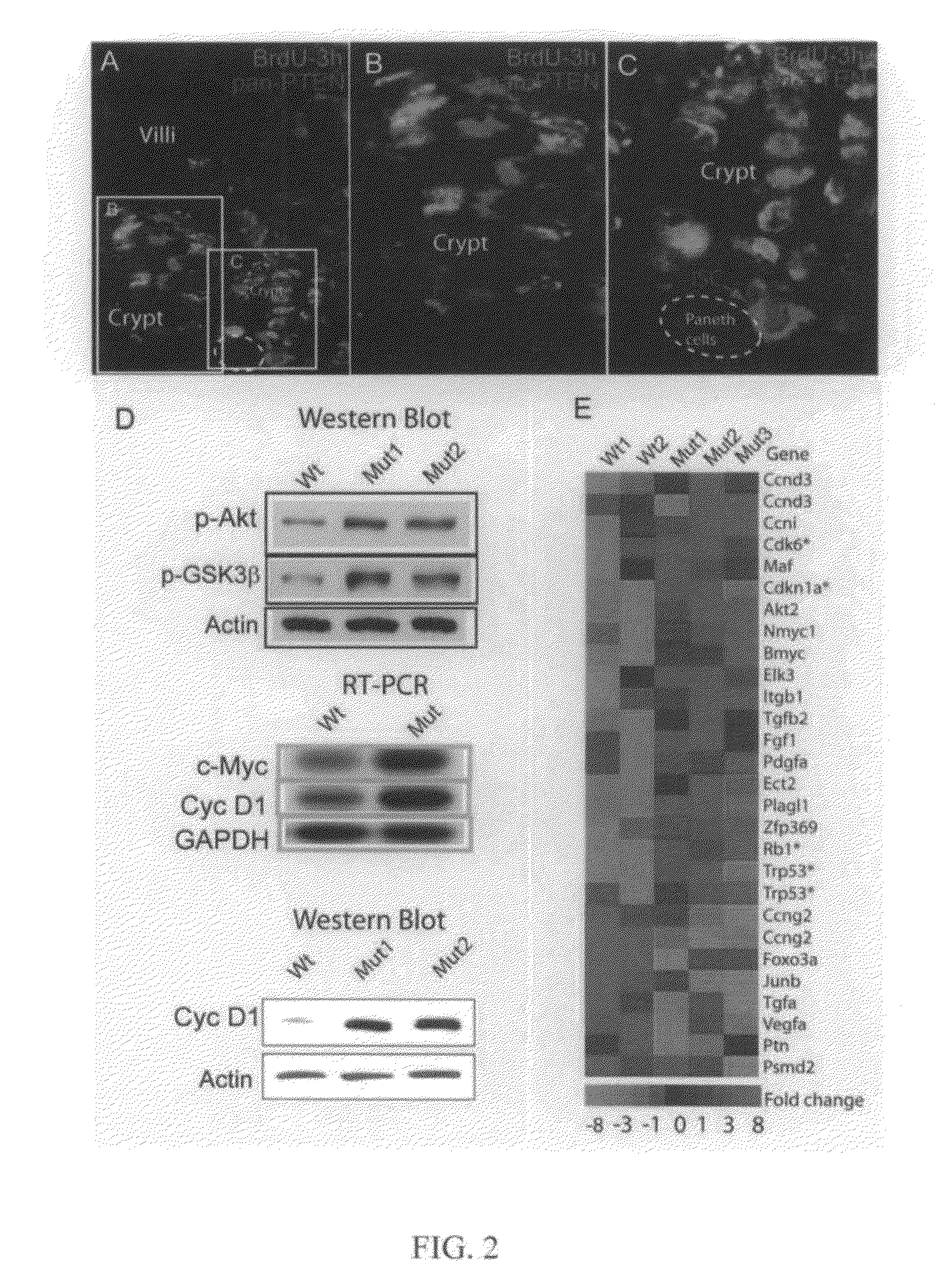
[0152]Next, the relationship between PTEN expression and cell cycle status was examined. PTEN can be phosphorylated at its carboxy terminus by casein kinase II, which imp...
example 3
Musashi Positive Stem / Progenitor Cells Initiate Polyp Formation
[0155]The observations that PTEN is expressed in ISCs (FIG. 2C) and that PTEN loss results in polyposis raise the possibility that PTEN-deficient ISCs are responsible for the polyp formation. The ISC marker Musashi (Musashi1) was used to test this hypothesis. In normal intestinal crypts two classes of Musashi+ cells were detected by a polyclonal anti-Musashi1 antibody (Chemicon; Cat # AB5977). As expected, many Musashi+crypt cells were detected at the ISC position and could be co-stained for long-term-retention of BrdU (BrdU-LTR) (FIG. 3A), but some isolated Musashi+BrdU-LTR− cells (potentially progenitor cells) were observed higher in the crypt (FIG. 3A) and in the transition zone between crypt and villus (FIG. 8E). A rat monoclonal anti-Musashi1 antibody yielded a somewhat broader pattern of signal but had a similar distribution of strongly positive cells (Potten et al., 2003; FIG. 10). The differential ability of the ...
PUM
 Abstract
Description
Claims
Application Information
 - R&D
- Intellectual Property
- Life Sciences
- Materials
- Tech Scout
- Unparalleled Data Quality
- Higher Quality Content
- 60% Fewer Hallucinations
Browse by: Latest US Patents, China's latest patents, Technical Efficacy Thesaurus, Application Domain, Technology Topic, Popular Technical Reports.
© 2025 PatSnap. All rights reserved.Legal|Privacy policy|Modern Slavery Act Transparency Statement|Sitemap|About US| Contact US: help@patsnap.com


