
Intra-lumen polyp detection

a detection field and lumen technology, applied in the field of intralumen polyp detection, can solve the problems of poor prognosis, 50% or less 5-year survival rate, 30% recurrence rate,

Active Publication Date: 2007-07-12
CHECK CAP
View PDF70 Cites 61 Cited by
  • Summary
  • Abstract
  • Description
  • Claims
  • Application Information

AI Technical Summary

Benefits of technology

[0045] In an embodiment, the capsule includes an inflatable balloon, adapted to inflate around the capsule. For some applications, the balloon is configured such that the capsule moves towards a center of the balloon upon inflation thereof. For some applications, the balloon is configured to inflate when the capsule reaches an area of clinical interest within the GI tract. For some applications, the balloon includes a valve, adapted to open a certain period of time after the capsule reaches the area of clinical interest, thereby allowing the balloon to deflate.
[0051] In an embodiment, the capsule includes at least one radiation shield. For some applications, the at least one shield is configured to prevent radiation from being emitted from the radiation source in directions other than a single confined solid sector relative to a sphere surrounding the capsule.

Problems solved by technology

Clinical evidence suggests that early detection of primary colorectal cancer leads to a 90% or better 5-year survival rate, while detection of the disease when it has already metastasized leads to poor prognosis with a 50% or less 5-year survival rate and a 30% recurrence rate.

Method used


Image

  • Intra-lumen polyp detection
  • Intra-lumen polyp detection
  • Intra-lumen polyp detection

Examples


Embodiment Construction

[0148] FIGS. 2A-D are schematic illustrations of apparatus for conducting an exemplary experiment that illustrates physical principles upon which some embodiments of the present invention are based, in accordance with an embodiment of the present invention. A container 12 is filled with a radio-opaque contrast agent 10 in liquid or low viscosity gel, and a reservoir 17 is placed below the container and filled with water 11. A small water-filled balloon 18 is placed at the bottom of the container. In this experiment, container 12 filled with contrast agent 10 simulates a colon filled with contrast agent, water-filled reservoir 17 simulates tissues and organs outside the colon, and water-filed balloon 18 simulates an anatomical abnormality, such a polyp.

[0149] In the experiment, a radiation source 14 and a radiation detector 16 are placed near each other. The open ends of a collimator 13 for source 14 and a collimator 19 for detector 16 are facing the liquid container. Radiation sour...


PUM


Abstract

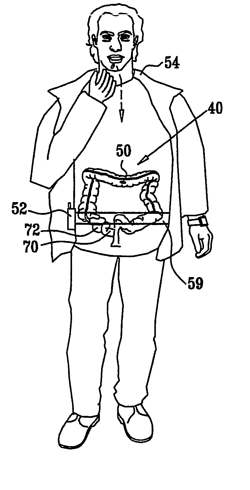
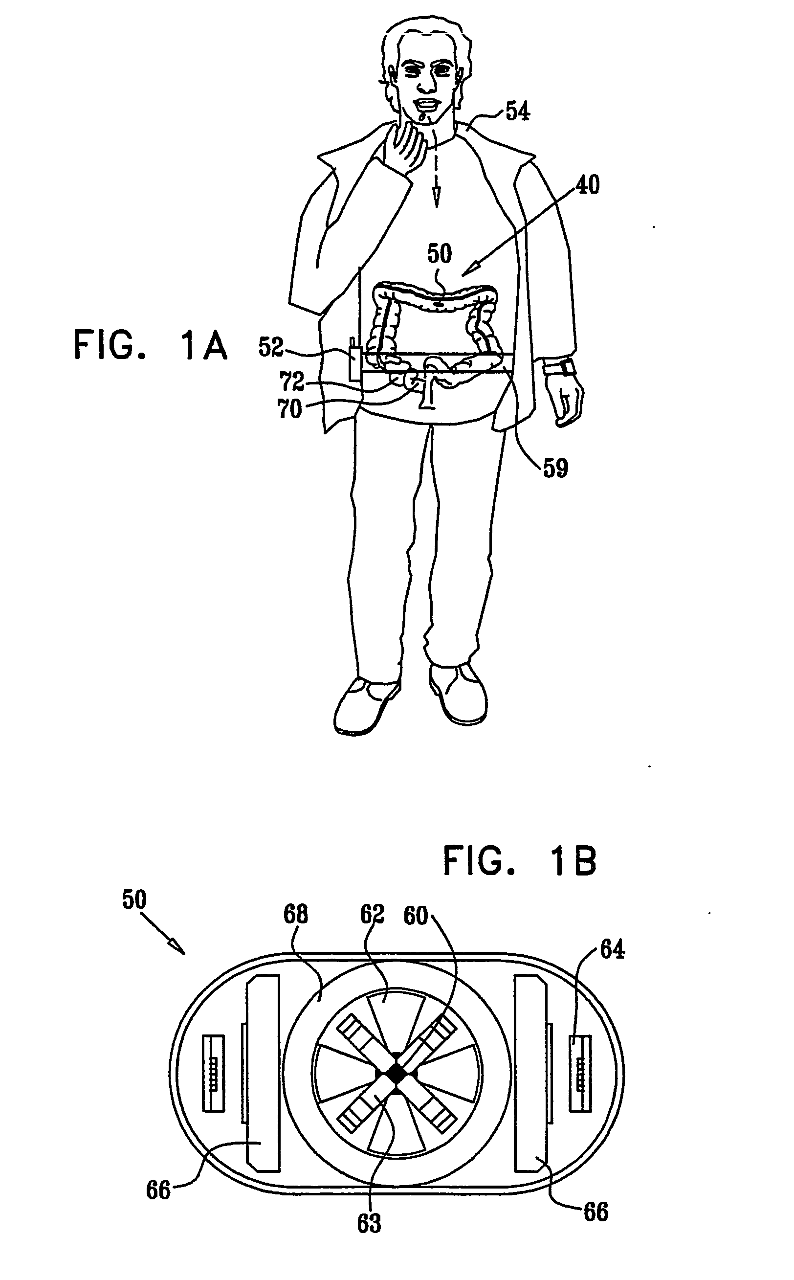
Apparatus (40) is provided, including a capsule (50), adapted to be swallowed by a subject (54), the capsule (50) including (a) at least one radiation source (60), adapted to emit radiation having an energy of at least 10 keV, and (b) at least one photon detector (62), adapted to detect photons generated responsively to the emitted radiation, the photons having an energy of at least 10 keV. The apparatus (40) additionally includes a control unit (64), adapted to analyze data regarding the photons in order to generate information useful for identifying a clinically-relevant feature of a gastrointestinal (GI) tract (72) of the subject (54).

Description

CROSS-REFERENCES TO RELATED APPLICATIONS [0001] This application claims the benefit of: (a) U.S. Provisional Patent Application 60 / 531,690, filed Dec. 17, 2003, entitled, “Intra lumen polyp detection,” and (b) U.S. Provisional Patent Application 60 / 559,695, filed Mar. 31, 2004, entitled, “Intra-lumen polyp detection.” Both of these application are assigned to the assignee of the present application and are incorporated herein by reference.FIELD OF THE INVENTION [0002] The present invention relates generally to the field of detection of conditions of a body lumen, and specifically to a swallowable device that travels in the colon and detects anatomical anomalies. BACKGROUND OF THE INVENTION [0003] Colorectal cancer is one of the leading causes of death in the Western world. Clinical evidence suggests that early detection of primary colorectal cancer leads to a 90% or better 5-year survival rate, while detection of the disease when it has already metastasized leads to poor prognosis w...

Claims


Application Information

Patent Timeline
Patent Type & Authority Applications(United States)
IPC IPC(8): A61B5/05A61BA61B5/07
CPCA61B5/073A61B5/42A61B6/4057A61B6/425A61B6/547A61B6/481A61B6/482A61B6/483A61B6/485A61B6/4258
Inventor KIMCHY, YOAV
Owner CHECK CAP
Learn More