


Quick Research
Generate reliable direction feasibility study reports for your R&D in just a few steps.

Technical Q&A
Discover and master advanced knowledge NOW. Basics, ideas, possibilities, all at once.

Find Solutions
As an expert in R&D theories, this can generate solutions to your technical problems instantly.

Evaluate Feasibility
Analyze your overall solution with one click, know your potential R&D risks in advance.

Monitor Landscape
Get weekly tech updates, stay abreast of the latest tech innovations and key insights.
Method for Predicting Response to Epidermal Growth Factor Receptor-Directed Therapy
a technology of epidermal growth factor and response prediction, applied in the field of methods for predicting the response to cancer therapy, can solve the problems of limited therapeutic benefit and often toxic therapies for patients
- Summary
- Abstract
- Description
- Claims
- Application Information
AI Technical Summary
Benefits of technology
Problems solved by technology
Method used
Image

Examples
example 1
Staining Procedure for Biomarkers
[0041] Human tumor tissue samples were stained as follows. Tumor tissue in 10% Neutral Buffered Formalin Paraffin blocks are sectioned at 4 microns and the sections placed onto coated slides. EGFR immunostaining is preformed by using Ventana Medical Instruments, Inc. monoclonal 111.6; HER-2 immunostaining is performed by using Ventana Medical Instruments, Inc. monoclonal CB11, and HER-3 immunostaining is performed by using Ventana Medical Instruments, Inc. monoclonal SGP1. Her-1, Her-2, and Her-3 are immunostained using, for example, the “BenchMark” (VMSI) with I-VIEW (VMSI) detection chemistry. pEGFR immunostaining is performed by using Chemicon monoclonal MB3052. p-ERK (1:100) is obtained from Cell Signaling Technology (Beverly, Mass.) and immunostained using a labeled streptavidin peroxidase technique.
[0042] For example, slides for p-ERK (1:100) are antigen retrieved using 0.1 M citrate buffer, pH 6.0 in the “decloaker” (Biocare Corp.) and the s...
example 2
Procedure for Immunohistochemistry
[0043] Quantitative immunohistochemistry (IHC) is performed as previously described (Bacus, S., et al., Analyt. Quant. Cytol. Histol. 19, 316-328 (1997)). EGFR, and erbB-2 immunostaining is performed using the pre-diluted EGFR (Ventana monoclonal 111.6) and erbB-2 (Ventana monoclonal CB11) antibodies from Ventana on the VMSI automated “BenchMark” staining module as described. The VMSI “I-View” detection kit is used for all of the VMSI pre-diluted primary antibodies. HER-3 is also immunostained using the “BenchMark” with I-VIEW detection chemistry. pErk is immunostained using a labeled streptavidin peroxidase technique. Phospho-Erk1 / 2 slides are antigen retrieved as described (Bacus, S., et al., Analyt. Quant Cytol. Histol. 19, 316-328 (1997)). Slides are placed onto the Autostainer (Dako Corp.) and the “LSAB2” kit (Dako) employed as the detection chemistry. pEGFR is immunostained in a similar labeled streptavidin peroxidase technique. pEGFR slides ...
example 3
Analysis of Treatment with an Epidermal Growth Factor-Directed Therapy
[0045] 53 samples from renal cancer patients enrolled in a clinical trial sponsored by Abgenix and Immunex Corporation for an investigational drug directed to EGFR were analyzed for expression of various biomarkers. The sample slides were obtained from Impath Laboratories, Inc.
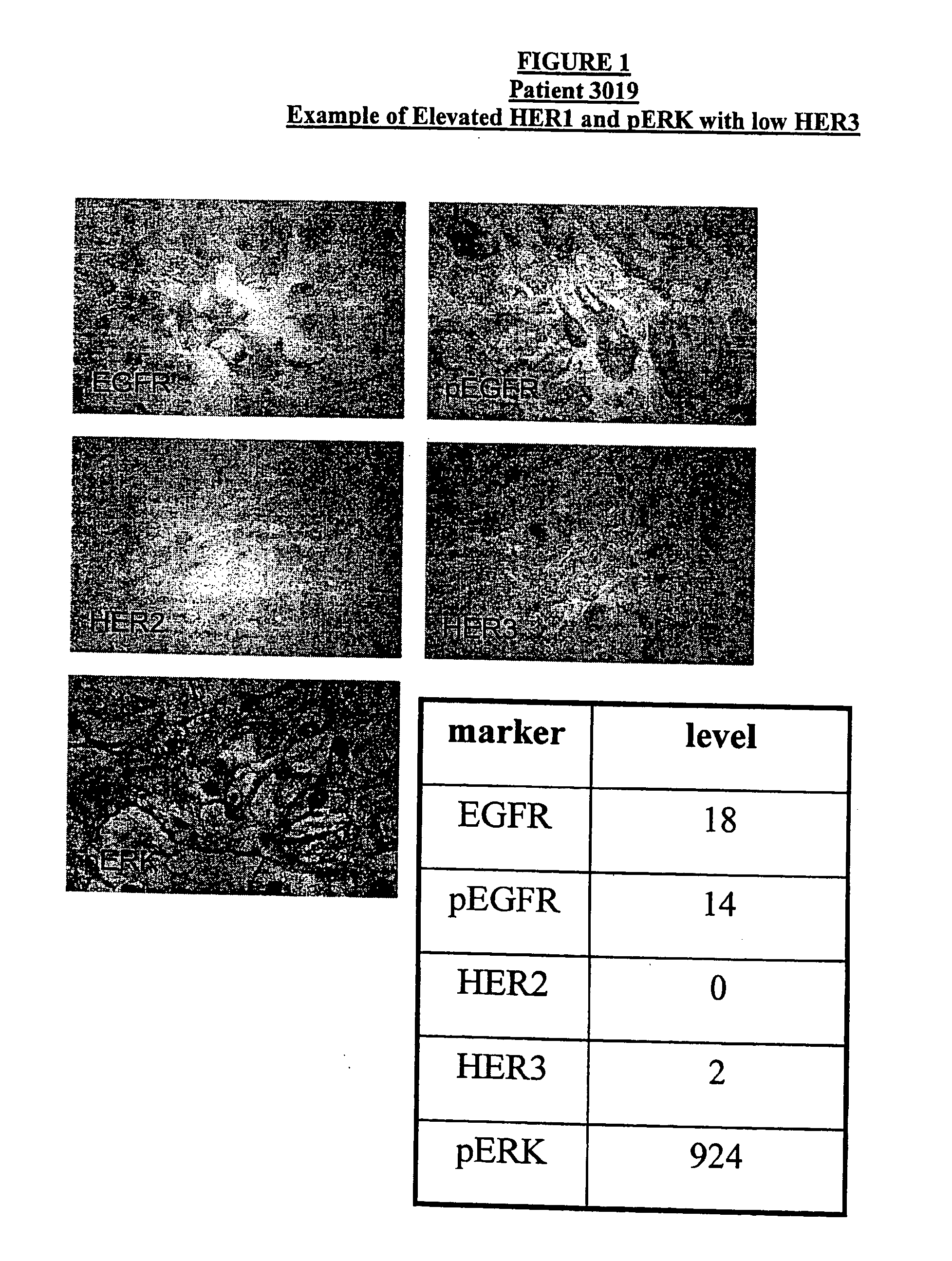
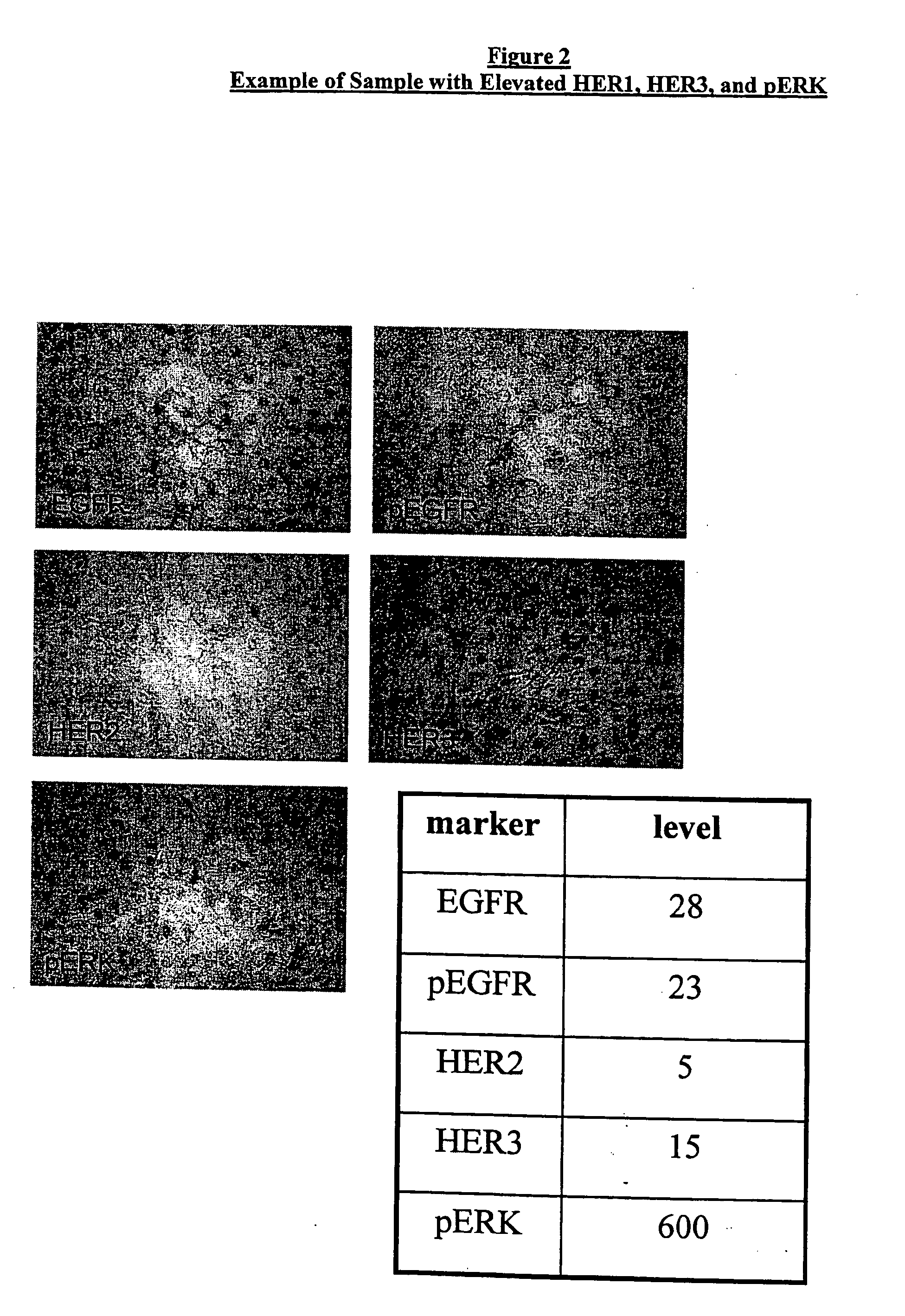
[0046] Immunohistochemical (IHC) analyses were carried out using the automated staining devices as described above. The antibodies used for the specific biomarkers included: Ventana monoclonal 111.6 for EGFR, Chemicon monoclonal MB3052 for pEGFR, polyclonal pERK from Cell Signaling Technology for pERK, Ventana monoclonal SGP1 for HER3, and Ventana monoclonal CB11 for HER2. For each specimen, a slide was stained with control mouse immunoglobulins to establish the existence and localization of background staining. In addition, appropriate positive controls were run for each IHC stain. Following counterstaining with ethyl green, the slides we...
PUM
| Property | Measurement | Unit |
|---|---|---|
| Volume | aaaaa | aaaaa |
| Volume | aaaaa | aaaaa |
| Volume | aaaaa | aaaaa |
Abstract
Description
Claims
Application Information
 - R&D Engineer
- R&D Manager
- IP Professional
- Industry Leading Data Capabilities
- Powerful AI technology
- Patent DNA Extraction
Browse by: Latest US Patents, China's latest patents, Technical Efficacy Thesaurus, Application Domain, Technology Topic, Popular Technical Reports.
© 2024 PatSnap. All rights reserved.Legal|Privacy policy|Modern Slavery Act Transparency Statement|Sitemap|About US| Contact US: help@patsnap.com