
System and Method For Myocardium Segmentation In Realtime Cardiac MR Data

Inactive Publication Date: 2007-05-24
SIEMENS MEDICAL SOLUTIONS USA INC
View PDF4 Cites 32 Cited by
  • Summary
  • Abstract
  • Description
  • Claims
  • Application Information

AI Technical Summary

Problems solved by technology

Although a physician can visually detect abnormalities of wall motions, mere interpretation of wall motion has well known limits.
For instance, a human cannot identify transient changes in myocardial motion, even if real-time MR provides high frame rate data acquisition.
However, real-time MR usually has low spatial resolution, thus reducing the amount of data to be processed.
On the other hand, the image data is noisy and less accurate.

Method used


Image

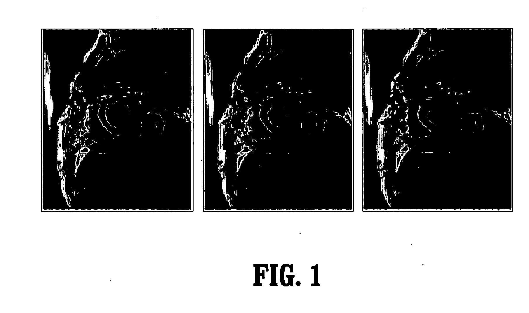
  • System and Method For Myocardium Segmentation In Realtime Cardiac MR Data
  • System and Method For Myocardium Segmentation In Realtime Cardiac MR Data
  • System and Method For Myocardium Segmentation In Realtime Cardiac MR Data

Examples


Embodiment Construction

[0022] Exemplary embodiments of the invention as described herein generally include systems and methods for fast segmentation of the myocardium. Accordingly, while the invention is susceptible to various modifications and alternative forms, specific embodiments thereof are shown by way of example in the drawings and will herein be described in detail. It should be understood, however, that there is no intent to limit the invention to the particular forms disclosed, but on the contrary, the invention is to cover all modifications, equivalents, and alternatives falling, within the spirit and scope of the invention.

[0023] As used herein, the term “image” refers to multi-dimensional data composed of discrete image elements (e.g., pixels for 2-D images and voxels for 3-D images). The image may be, for example, a medical image of a subject collected by computer tomography, magnetic resonance imaging, ultrasound, or any other medical imaging system known to one of skill in the art. The im...


PUM


Abstract

A method for cardiac segmentation includes providing a plurality of digitized medical images, each image comprising a plurality of intensities corresponding to a domain of points on an 2-dimensional grid, the plurality of images forming a sequence of cardiac images acquired at different time points, filtering noise from the sequence of images, approximating the endocardium with a first circle, approximating the epicardium with a second circle, finding candidate points for the endocardium, finding candidate points for the epicardium, and interpolating gaps between points of the endocardium and between points of the epicardium wherein the myocardium is segmented.

Description

CROSS REFERENCE TO RELATED UNITED STATES APPLICATIONS [0001] This application claims priority from: “A Fast Algorithm for the Segmentation of Myocardium in Realtime Cardiac MR Data”, U.S. Provisional Application No. 60 / 727,370 of Hong Shen, filed Oct. 17, 2005, the contents of which are incorporated herein by reference.TECHNICAL FIELD [0002] This invention is directed to cardiac segmentation in digitized medical images. DISCUSSION OF THE RELATED ART [0003] Cardiac magnetic resonance (“MR”) imaging has become a mainstream clinical instrument to assess congenital heart disease. It is a cutting-edge technology that is beginning to replace fluoroscopy as the modality of choice to guide minimally invasive interventions in the heart. [0004] Although a physician can visually detect abnormalities of wall motions, mere interpretation of wall motion has well known limits. For instance, a human cannot identify transient changes in myocardial motion, even if real-time MR provides high frame rat...

Claims


Application Information

Patent Timeline
IPC IPC(8): G06K9/00G06K9/34G06K9/36
CPCG06T7/0083G06T2207/10088G06T2207/20061G06T2207/30048G06T7/12
Inventor SHEN, HONG
Owner SIEMENS MEDICAL SOLUTIONS USA INC
Learn More