
Method and Apparatus for Non-Invasive Monitoring of Blood Substances Using Self-Sampled Tears

a blood substance and self-sampling technology, applied in the field of non-invasive monitoring of blood substances, can solve the problems of limited patient compliance with self-testing, three-fold increase in hypoglycemic incidents, and disability and death

Inactive Publication Date: 2007-02-22
EYELAB GROUP
View PDF9 Cites 15 Cited by
  • Summary
  • Abstract
  • Description
  • Claims
  • Application Information

AI Technical Summary

Benefits of technology

[0011] Therefore, it is an object according to the present invention to provide a method and apparatus for determining the concentration of a substance in tears which will allow for indirect monitoring of the substance concentration in blood.
[0012] It is a further object according to the present invention to provide an improved method and apparatus for non-invasively determining blood glucose concentration in a simple and accurate manner.

Problems solved by technology

Diabetes is a leading cause of disability and death, affecting approximately seventeen million Americans.
However, the trial resulted in a three-fold increase in hypoglycemic incidents.
This procedure is painful and inconvenient for even the most compliant patients, such that limited patient compliance with self-testing is a significant problem in the medical management of this disease.
Some of these methods have worked well in controlled laboratory testing, but in practice other chemical species, tissue optics, variations in temperature, and other factors have confounded the measurement.
For all spectroscopic approaches, the major problem is the need for frequent calibration, as infrared absorption bands for various chemicals in blood or interstitial fluid can overlap significantly and are influenced by temperature and hydrogen bonding effects.
In the aforementioned Chen study, glucose concentration in microliter samples of human tears obtained with capillary tubes was determined by capillary electrophoresis (CE) with laser-induced fluorescence (LIF), a sophisticated method limited to research chemistry laboratories because of its technical complexity.
Currently, no practical, entirely non-invasive system and method exists for patients to self-monitor their blood glucose with the level of accuracy and responsiveness required.

Method used


Image

  • Method and Apparatus for Non-Invasive Monitoring of Blood Substances Using Self-Sampled Tears
  • Method and Apparatus for Non-Invasive Monitoring of Blood Substances Using Self-Sampled Tears
  • Method and Apparatus for Non-Invasive Monitoring of Blood Substances Using Self-Sampled Tears

Examples


Embodiment Construction

)

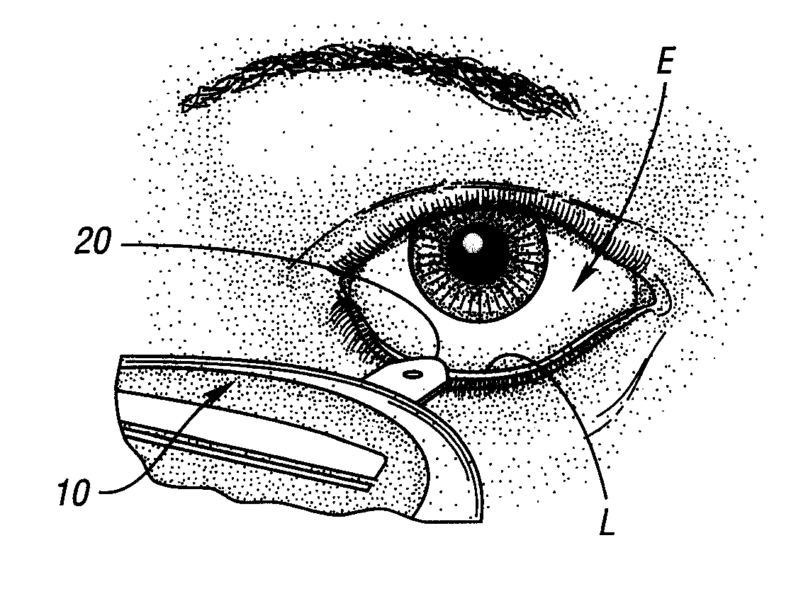
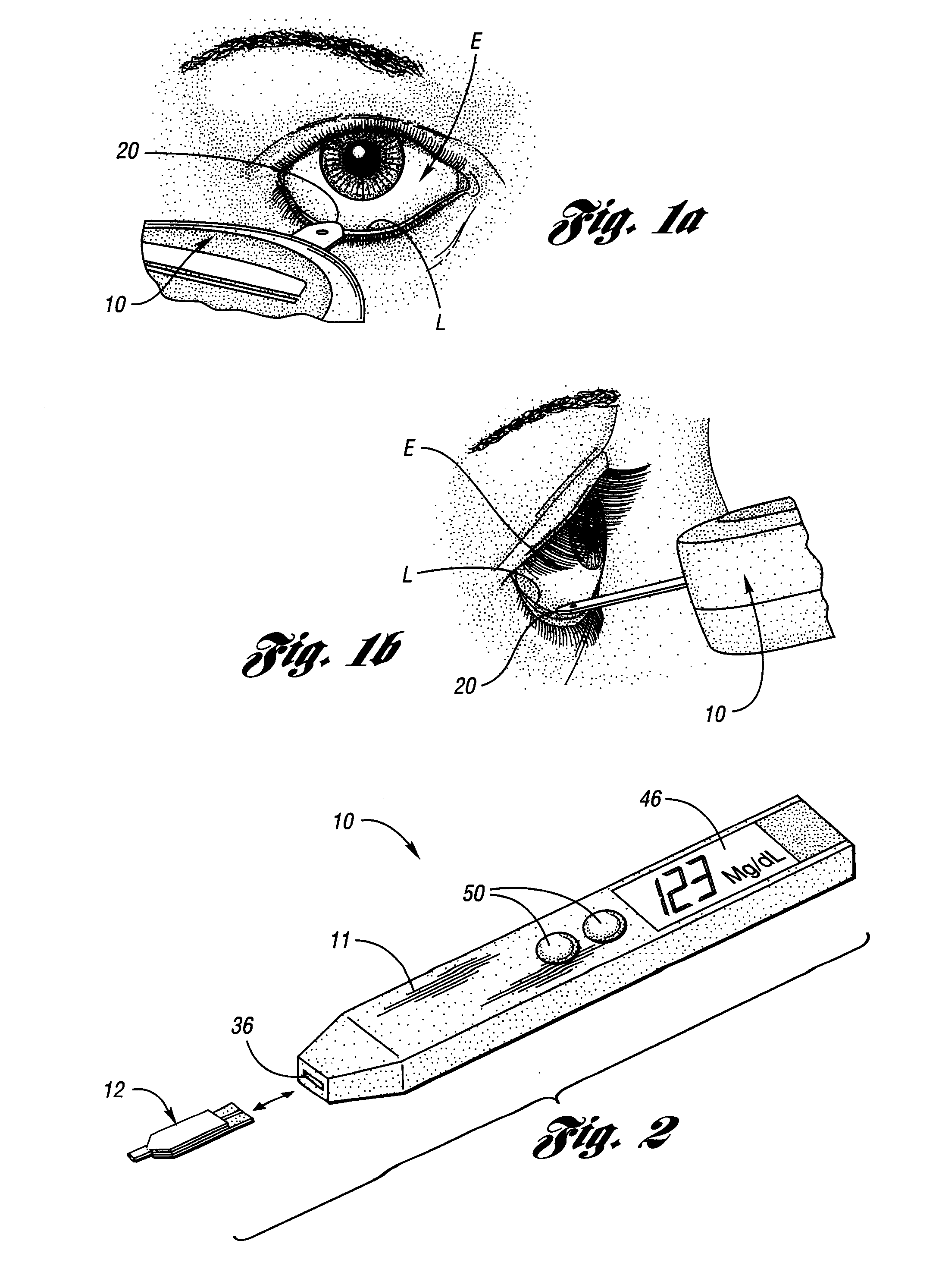
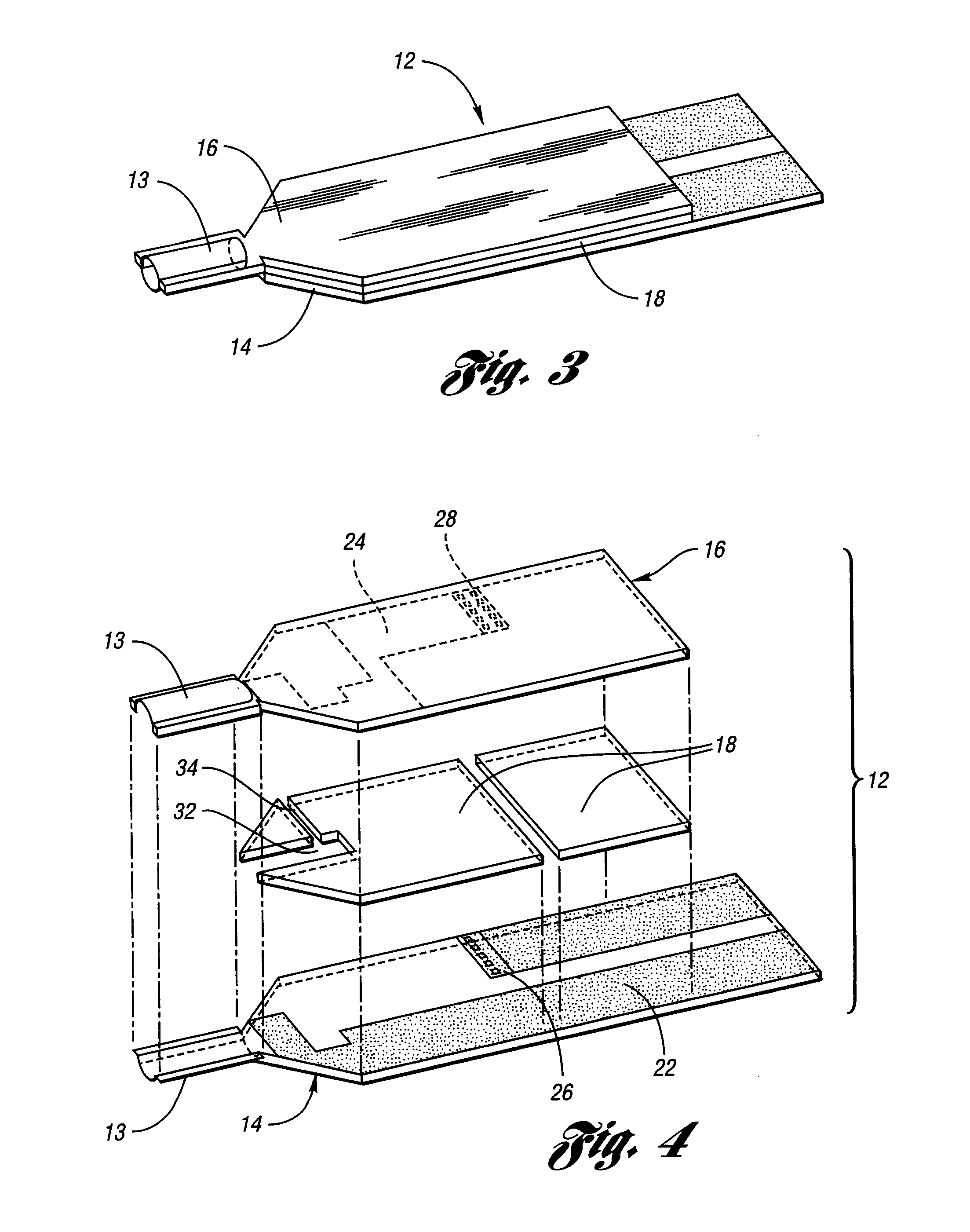
[0037] The method and apparatus of the present invention provide for the practical, non-invasive determination of the concentrations of substances, particularly glucose, in human tears in order to indirectly monitor the level of this important analyte in blood. The method and apparatus described herein are designed for the special limitations of analysis of tear fluid, namely the low glucose concentration in tears compared with blood and the small sample volume available. The present invention advances tight glycemic control of diabetes by permitting users to monitor their blood glucose levels by self-measuring the glucose levels in their tears, wherein the tear fluid sample is easily obtained by a user and accurate results are immediately available.

[0038] By way of background, the primary aqueous component of tears is secreted by the lacrimal gland, which is located beneath the outer portion of the upper eyelid. In this gland, a fraction of the glucose in blood crosses into the t...


PUM

PropertyMeasurementUnit
concentrationsaaaaaaaaaa
volumeaaaaaaaaaa

Abstract

A method and apparatus for non-invasively determining the concentration of a substance in blood, such as glucose, include a sample portion arranged for contacting an eye region of a user to obtain a tear fluid sample, a sensor in communication with the sample portion for generating a signal related to the tear substance concentration, and a processor in communication with the sensor for determining a blood substance concentration corresponding to the tear substance concentration.

Description

CROSS-REFERENCE TO RELATED APPLICATIONS [0001] This application is a continuation of U.S. application Ser. No. 10 / 404,702 filed Apr. 1, 2003 which, in turn, claims the benefit of U.S. provisional application Ser. No. 60 / 370,552 filed Apr. 5, 2002.BACKGROUND OF THE INVENTION [0002] 1. Field of the Invention [0003] This invention relates to a method and apparatus for non-invasive monitoring of blood substances, particularly glucose, using self-sampled tears. [0004] 2. Background Art [0005] The measurement of glucose in blood plasma is perhaps the most important physiologic analyte measurement in medicine, as diabetes has immense public health implications. Diabetes is a leading cause of disability and death, affecting approximately seventeen million Americans. The total annual cost of treating diabetes and its complications in the United States is in excess of $150 billion, a large part of the total national expenditure for health care. [0006] The medical management of diabetes by tig...

Claims


Application Information

Patent Timeline
Patent Type & Authority Applications(United States)
IPC IPC(8): A61B5/05A61B5/00C12Q1/00C12Q1/32C12Q1/54G01N1/10G01N1/14G01N33/487G01N33/52G01N35/04
CPCA61B5/1486A61B2562/0295C12Q1/006C12Q1/32C12Q1/54G01N27/3272G01N33/528G01N2001/1056G01N2001/149G01N2035/0465Y10T436/144444
Inventor COHAN, BRUCE E.CHA, GEUN SIGMEYERHOFF, MARK E.NAM, HAKHYUNGILLESPIE, DONALD E.CUI, GANGKIM, JONG SIKFLANDERS, ZVI
Owner EYELAB GROUP
Learn More