
Method and system for automated digital image analysis of prostrate neoplasms using morphologic patterns

a prostrate neoplasm and digital image technology, applied in image analysis, image enhancement, instruments, etc., can solve the problems of lack of national awareness and research funding breast cancer currently receives, inability to confirm the presence of prostate cancer by itself, and difficulty in diagnosing prostatic adenocarcinoma, especially when present in small amounts

Inactive Publication Date: 2007-01-25
VENTANA MEDICAL SYST INC
View PDF4 Cites 46 Cited by
  • Summary
  • Abstract
  • Description
  • Claims
  • Application Information

AI Technical Summary

Problems solved by technology

Prostate cancer strikes as many men (and causes almost as many deaths annually) as breast cancer does in women, but lacks the national awareness and research funding breast cancer currently receives.
But since other kinds of prostate disease can also cause high PSA levels, PSA testing by itself cannot confirm the presence of prostate cancer.
Moreover, the diagnosis of prostatic adenocarcinoma, especially when present in small amounts, is often challenging.

Method used


Image

  • Method and system for automated digital image analysis of prostrate neoplasms using morphologic patterns
  • Method and system for automated digital image analysis of prostrate neoplasms using morphologic patterns
  • Method and system for automated digital image analysis of prostrate neoplasms using morphologic patterns

Examples


Embodiment Construction

Exemplary Biological Sample Analysis System

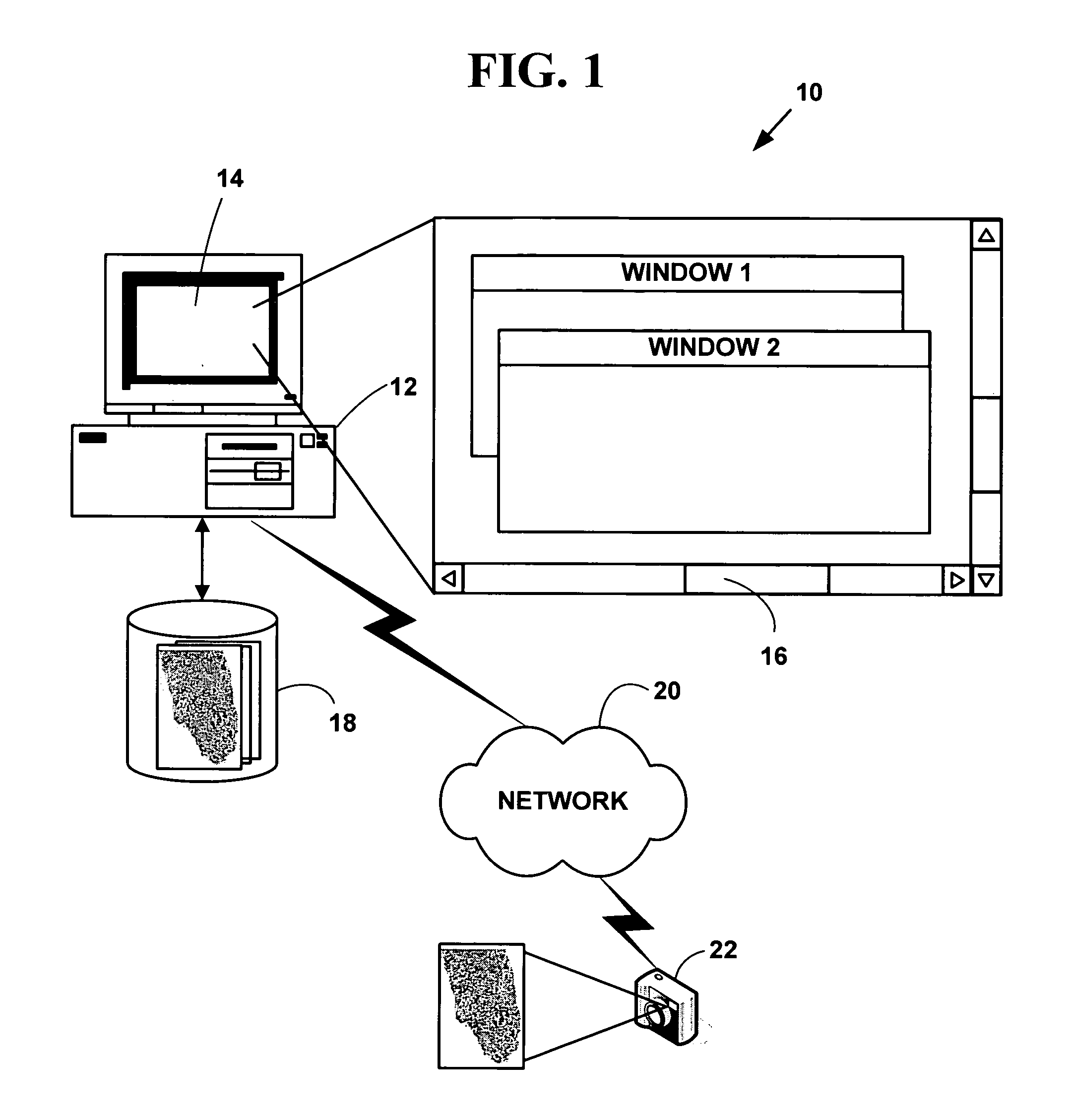
[0044]FIG. 1 is a block diagram illustrating an exemplary biological sample analysis processing system 10. The exemplary biological sample analysis processing system 10 includes one or more computers 12 with a computer display 14 (one of which is illustrated). The computer display 14 presents a windowed graphical user interface (“GUI”) 16 with multiple windows to a user. The present invention may optionally include a microscope or other magnifying device (not illustrated in FIG. 1) and / or a digital camera 18 or analog camera. One or more databases 20 (one or which is illustrated) include biological sample information in various digital images or digital data formats. The databases 20 may be integral to a memory system on the computer 12 or in secondary storage such as a hard disk, floppy disk, optical disk, or other non-volatile mass storage devices. The computer 12 and the databases 20 may also be connected to an accessible via one or mo...


PUM


Abstract

A method and system method and system automated digital image analysis of prostrate neoplasms using morphologic patterns. The method and system provide automated screening of prostate needle biopsy specimens in a digital image and automated diagnosis of prostatectomy specimens.

Description

CROSS REFERENCES TO RELATED APPLICATIONS [0001] This application claims priority to U.S. Provisional Patent Application No. 60 / 679,449, filed May 10, 2005, and U.S. patent application Ser. No. 11 / 361,774, filed Feb. 23, 2006, which claims priority to U.S. Provisional Patent Application No. 60 / 655,465, filed Feb. 23, 2005, the contents of all of which are incorporated by reference.COPYRIGHT NOTICE [0002] Pursuant to 37 C.F.R. 1.71(e), applicants note that a portion of this disclosure contains material that is subject to and for which is claimed copyright protection, such as, but not limited to, digital photographs, screen shots, user interfaces, or any other aspects of this submission for which copyright protection is or may be available in any jurisdiction. The copyright owner has no objection to the facsimile reproduction by anyone of the patent document or patent disclosure, as it appears in the U.S. Patent Office patent file or records. All other rights are reserved, and all othe...

Claims


Application Information

Patent Timeline
Patent Type & Authority Applications(United States)
IPC IPC(8): G06K9/00
CPCG06T2207/30004G06T7/0012
Inventor GHOLAP, ABHIJEET S.NAIK, GAURI A.JOSHI, APARNASAWAIMOON, SATYAKAMSIDDHESHWAR, CHIVATE SUJITJADHAV, PRITHVIRAJRAO, C. V. K.
Owner VENTANA MEDICAL SYST INC
Learn More