
Methods and apparatus to achieve a closure of a layered tissue defect

Inactive Publication Date: 2006-11-30
TERUMO KK
View PDF47 Cites 300 Cited by
  • Summary
  • Abstract
  • Description
  • Claims
  • Application Information

AI Technical Summary

Benefits of technology

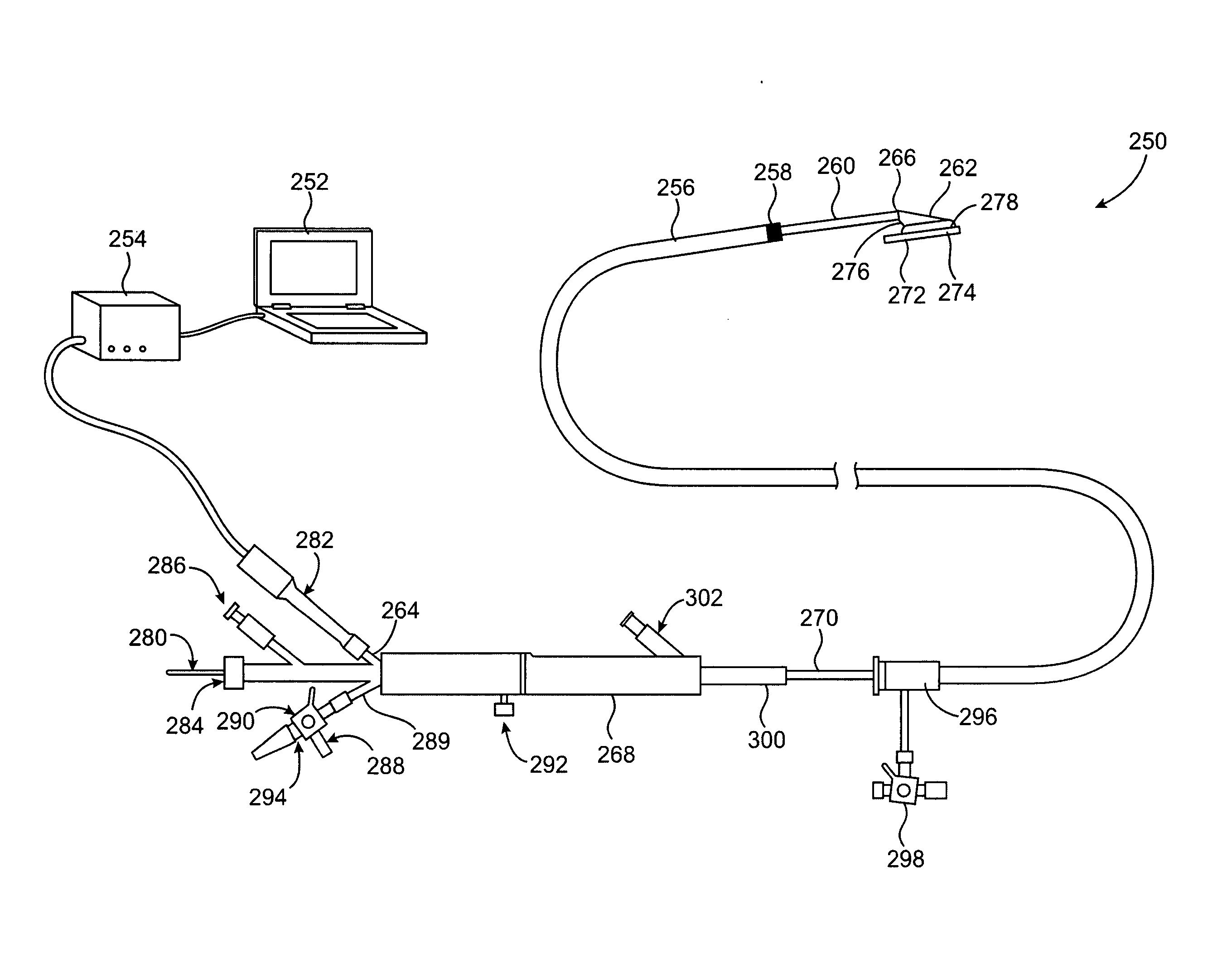
[0029] In another aspect of the present invention, a system for fusing layered tissue structures comprises a catheter body having a proximal and distal end, a housing on a distal portion of the catheter body, an introducer sheath having a main body as well as proximal and distal ends, that is slidably disposed over a portion of the catheter body and an energy transmission member positioned within the housing. The energy transmission member and the housing are collapsible and slidably movable relative to the introducer sheath from a collapsed position within the introducer sheath to an expanded position beyond the distal end of the introducer sheath. Preferably, the introducer sheath has a softer durometer distal tip than the main body of the introducer sheath and this tip facilitates movement of the housing and the energy transmission member from the expanded position to the collapsed position within the introducer sheath. The softer durometer tip may be integral with the main body or it may be fixedly connected to the main body.
[0030] In the system, the introducer sheath may comprise a valve adapted to accommodate the housing and this valve also minimizes blood loss from the introducer sheath. Typically the valve is a hemostasis valve which may include one or more valve membranes such as disks which have a top surface and a bottom surface, both of which are scored. They may be scored orthogonally or at other angles.
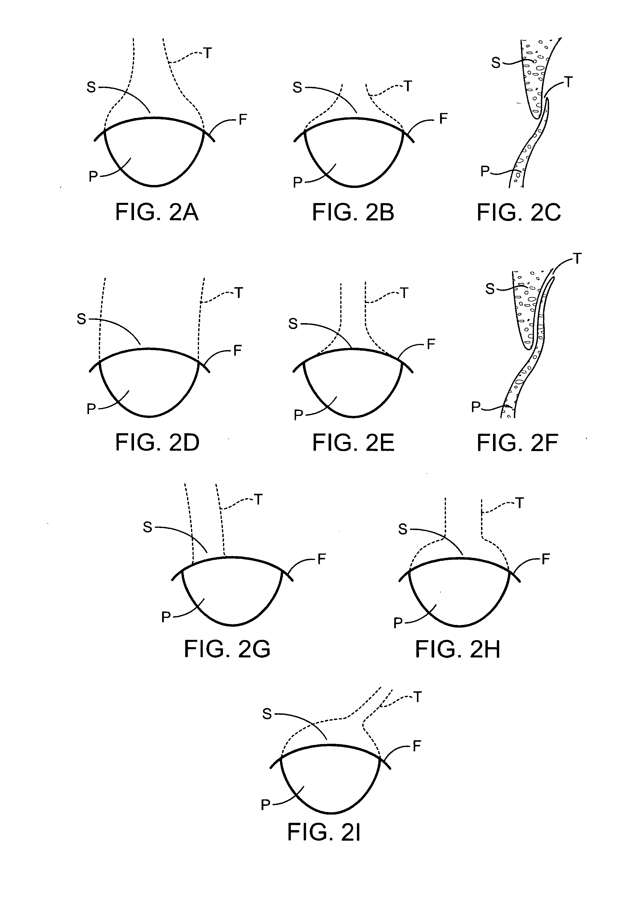
[0032] In still another aspect of the present invention, a method for fusing apposed layered tissue structures comprises positioning a closure device at a first treatment site having a first layer of tissue as well as a second layer to tissue. The layers of tissue are approximated and energy is applied from the closure device to the tissue thereby fusing the layers of tissue. The method can also include electrophysiological monitoring of the layered tissue as well as adjacent tissue so that creation of aberrant conductive pathways is minimized. This can be accomplished by minimizing delivery of energy as well as minimizing the surface area of the active electrode and / or distance of the treatment zone to the AV node of a patient's heart.

Problems solved by technology

It has been estimated that patients with PFOs who have already had cryptogenic strokes may have a risk of having another stroke.
The cost and potential side-effects and complications of such a procedure must be low, however, since the event rate due to PFOs is relatively low.
Currently available interventional therapies for defect closure are generally fairly invasive and / or have potential drawbacks.
Performing open heart surgery purely to close an asymptomatic PFO or even a very small ASD, however, would be very hard to justify.
Although available devices may work well in some cases, they also face a number of challenges.
Relatively frequent causes of complications include, for example, improper deployment, device embolization into the circulation and device breakage.
In some instances, a deployed device does not heal into the septal wall completely, leaving an exposed tissue which may itself be a nidus for thrombus formation.
Furthermore, currently available devices are generally complex and expensive to manufacture, making their use for prophylactic treatment of PFO and other defects impractical.
None of these disclosures, however, show methods or apparatus suitable for positioning the tissues of an anatomic defect for welding or for delivering the energy to an anatomic defect to be welded.
These disclosures do not teach methods that would be particularly useful for welding layered tissue structures such as PFOs, nor do they teach bringing together tissues of a defect such that a tissue overlap is created that can then be welded together.
Frequently, scar tissue may fail to form or may form incompletely, resulting in a still patent PFO.
In addition to PFO, a number of other anatomic tissue defects, such as other ASDs, ventricular septal defects (VSDs), patent ductus arteriosis (PDA), aneurysms and other blood vessel wall defects, atrial appendages and other naturally occurring cavities within which blood clots can form, and the like cause a number of different health problems (note that the term “defect” may include a naturally occurring structure that results a potential health risk such as the clot forming in the atrial appendage).

Method used


Image

  • Methods and apparatus to achieve a closure of a layered tissue defect
  • Methods and apparatus to achieve a closure of a layered tissue defect
  • Methods and apparatus to achieve a closure of a layered tissue defect

Examples


Embodiment Construction

[0088] Devices, systems, and methods of the present invention generally provide for treatment of anatomic defects in human tissue, such as a patent foramen ovale (PFO), atrial septal defect (ASD), ventricular septal defect (VSD), left atrial appendage (LAA), patent ductus arteriosis (PDA), vessel wall defects and / or the like through application of energy. The present invention is particularly useful for treating and fusing layered tissue structures where one layer of tissue at least partly overlaps a second layer of tissue as found in a PFO. Therefore, although the following descriptions and the referenced drawing figures focus primarily on treatment of PFO, any other suitable tissue defects, such as but not limited to those just listed, may be treated in various embodiments.

[0089] I. PFO Anatomy

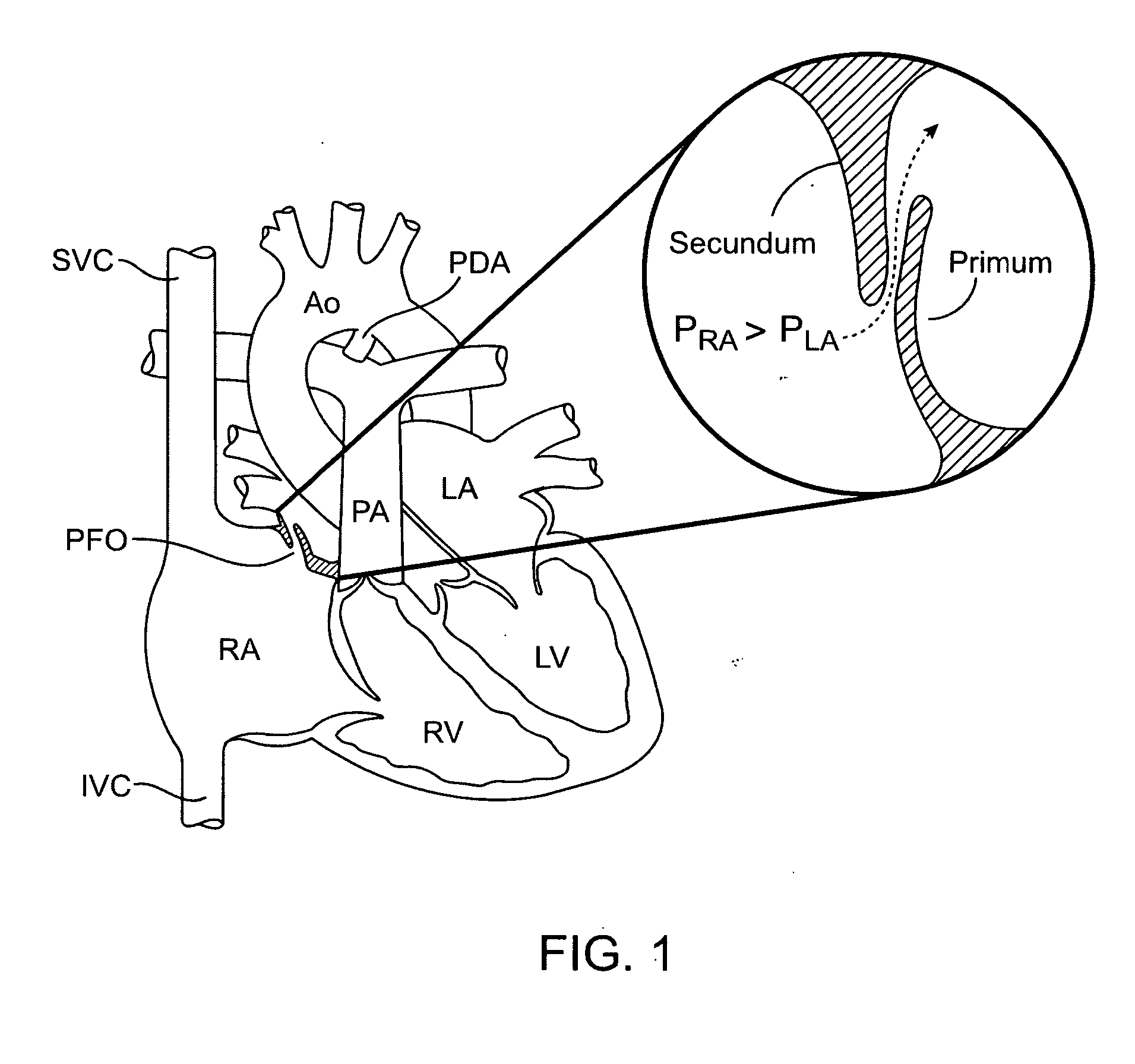
[0090] As mentioned in the background section above, FIG. 1 is a diagram of the fetal circulation. The foramen ovale is shown PFO, with an arrow expanded view demonstrating that blood pass...


PUM


Abstract

Methods for treating anatomic tissue defects such as a patent foramen ovate generally involve positioning a distal end of a catheter device at the site of the defect, exposing a housing and energy transmission member from the distal end of the catheter, engaging the housing with tissues at the site of the defect, applying suction or other approximating tool to the tissue via the housing to bring the tissue together, and applying energy to the tissue with the energy transmission member or to deliver a clip or fixation device to substantially close the defect. Apparatus generally include a catheter body, a housing extending from a distal end of the catheter body for engaging tissue at the site of the defect, and further adapted to house a fusing or fixation device such as an energy transmission member adjacent a distal end of the housing, or a clip or fixation delivery element.

Description

CROSS-REFERENCES TO RELATED APPLICATIONS [0001] The present application is a non-provisional of U.S. patent application Ser. No. 60 / 670,535 (Attorney Docket No. 022128-000700US), filed Apr. 11, 2005, the full disclosure of which is incorporated herein by reference.BACKGROUND OF THE INVENTION [0002] The invention generally relates to medical devices and methods. More specifically, the invention relates to positioning closure devices, including energy based devices and methods for treatment of anatomic defects in human tissue, such as a patent foramen ovale (PFO), atrial septal defect (ASD), ventricular septal defect (VSD), patent ductus arteriosis (PDA), left atrial appendages (LAA), blood vessel wall defects and other defects having layered and apposed tissue structures. [0003] The following is an example of how one particular type of anatomical defect, a PFO, is formed. Fetal blood circulation is very different from adult circulation. Because fetal blood is oxygenated by the placen...

Claims


Application Information

Patent Timeline
IPC IPC(8): A61B18/04A61B18/18
CPCA61B17/0057A61B2018/1407A61B2017/00504A61B2017/00557A61B2017/00561A61B2017/00575A61B2017/22069A61B2018/00351A61B2018/0063A61B2019/461A61M25/0069A61M25/10A61M2025/105A61M2025/1052A61M2025/1054A61B18/1492A61B2090/061
Inventor FRANCIS, DANALEJANDRO, JOSEENGELSON, ERIKFILLOUX, DOMINIQUEHORNE, KENNETHKIM, LUCIAKUMAR, UDAY N.SUTTON, DOUGTAIMISTO, MIRIAM H.UCHIDA, ANDY
Owner TERUMO KK
Learn More