
Methods and devices for anchoring to soft tissue

Inactive Publication Date: 2006-11-16
BOSTON SCI SCIMED INC +1
View PDF19 Cites 119 Cited by
  • Summary
  • Abstract
  • Description
  • Claims
  • Application Information

AI Technical Summary

Benefits of technology

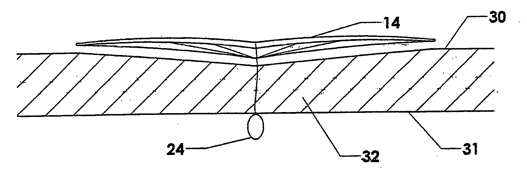
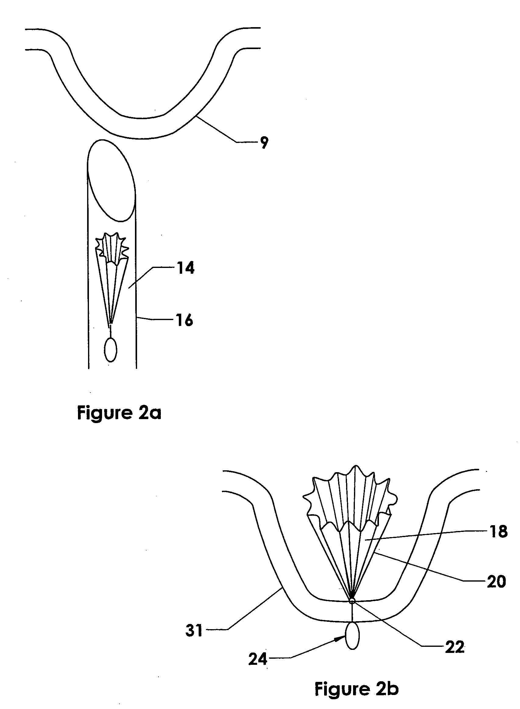
[0010] In the preferred embodiments, the anchoring elements can be delivered endoscopically, as through a rigid endoscope, or endoluminally, as through a flexible endoscope. The anchors are designed to collapse to a small diameter so that they can be placed into the working channel of an endoscope and then expand to a larger diameter upon deployment. The expanded diameter presents a large surface area and is designed such that any force applied to the anchor is distributed to this surface area and the resultant force per unit area is reduced as compared to sutures or staples. The resultant force per unit area is intended to be small enough to prevent pull-out of the anchor through the soft tissue wall.
[0021] An additional embodiment of the present invention utilizes a piece of mesh material that is deployed and spread out once through the soft tissue. The device utilizes supporting elements that move from a collapsed state to an uncollapsed or deployed state. In the deployed condition the supporting elements lock into position to provide structural integrity to the mesh. The mesh in all these embodiments may promote cell ingrowth or may facilitate attachment of cellular structures once ingrowth occurs.

Problems solved by technology

However, when segments of tissue are attached together and then exposed to tension post-operatively, such techniques often do not hold up over time.
The use of pledgets is not always possible especially when securing the wall of an organ that has a surface not easily accessible during the procedure.
Sutures that are placed through the wall can be strain-relieved with a pledget or similar device only along the inner surface of the wall, but not along the outer wall (unless a pledget or similar device is passed through the wall, which is generally not practical).
When sutures placed in this way are exposed to tension, as is the case when a gastroplasty procedure is done to create a gastric restriction, the sutures generally pull out over time.
Similarly, when attaching a foreign body to a segment of soft tissue using attachment techniques such as suturing, if the foreign body is subjected to forces postoperatively, the foreign body will typically pull loose from the tissue segment.

Method used


Image

  • Methods and devices for anchoring to soft tissue
  • Methods and devices for anchoring to soft tissue
  • Methods and devices for anchoring to soft tissue

Examples


Embodiment Construction

[0064] As has been described, the attachment of fastening devices to soft tissue often depends on the penetration of soft tissue and placement of pledgets, mesh, umbrellas or other anchors on the outer wall of soft tissue. This can be dangerous because other important blood vessels, nerves or organs such as the liver, lungs, heart, gall bladder, kidneys, reproductive organs or other sensitive tissue often reside close to the point of placement, and the exact location of these sensitive structures is rarely known prior to intervention on the soft tissue by the physician. Many of the methods and devices described in this application can be placed with the use of a “safe harbor” delivery system that permits penetration of the soft tissue wall and placement of the anchor on the opposite side with less fear of damage to surrounding sensitive structures.

[0065] As shown in FIG. 1a, an endoscope 1 with a working channel 2, is employed that has a stabilizing element 4 attached to the distal...


PUM


Abstract

Disclosed are methods and devices for placing anchors into soft tissue such as the walls of the stomach. The anchoring device includes a tissue interface that is introduced in a collapsed configuration and which expands radially outward. The tissue interface includes a framework capable of distributing a load applied to the linkage element across the surface area of the tissue interface.

Description

CLAIM OF PRIORITY [0001] This application claims priority under 35 U.S.C. §119(e) to U.S. Provisional Application No. 60 / 659,445, filed Feb. 22, 2005, the entire contents of which are hereby expressly incorporated by reference.BACKGROUND [0002] 1. Field of the Invention [0003] The present invention relates to devices and methods for attaching soft tissues and in particular, to novel tissue anchoring elements. [0004] 2. Description of the Related Art [0005] The securement of soft tissue segments has traditionally been done using suturing or stapling devices. However, when segments of tissue are attached together and then exposed to tension post-operatively, such techniques often do not hold up over time. For example, when two segments of the stomach are sewn together the sutures that hold the segments together are in tension post-operatively. In order to prevent the sutures from pulling through the stomach wall over time, the sites where the sutures puncture the outer wall of the sto...

Claims


Application Information

Patent Timeline
IPC IPC(8): A61B17/08
CPCA61B17/00234A61B17/04A61B17/0469A61B17/0482A61B17/0487A61B2017/0417A61F2/0077A61B2017/0454A61B2017/0464A61B2017/0496A61B2017/06052A61B2017/306A61F2/0063A61B2017/0419
Inventor KELLEHER, BRIANYUREK, MATT
Owner BOSTON SCI SCIMED INC
Learn More