
Methods and apparatuses for noninvasive determinations of analytes

a non-invasive and analyte technology, applied in the field of methods and measurement of analytes, can solve the problems of system itself introducing tissue noise, changes in the optical properties of tissue can contribute to tissue noise, and no group has demonstrated a system that generates adequate non-invasive glucose measurements, etc., to achieve accurate non-invasive determination of tissue properties, discourage light collection, and encourage light collection. preferential

Inactive Publication Date: 2006-08-10
INLIGHT SOLUTIONS
View PDF11 Cites 52 Cited by
  • Summary
  • Abstract
  • Description
  • Claims
  • Application Information

AI Technical Summary

Benefits of technology

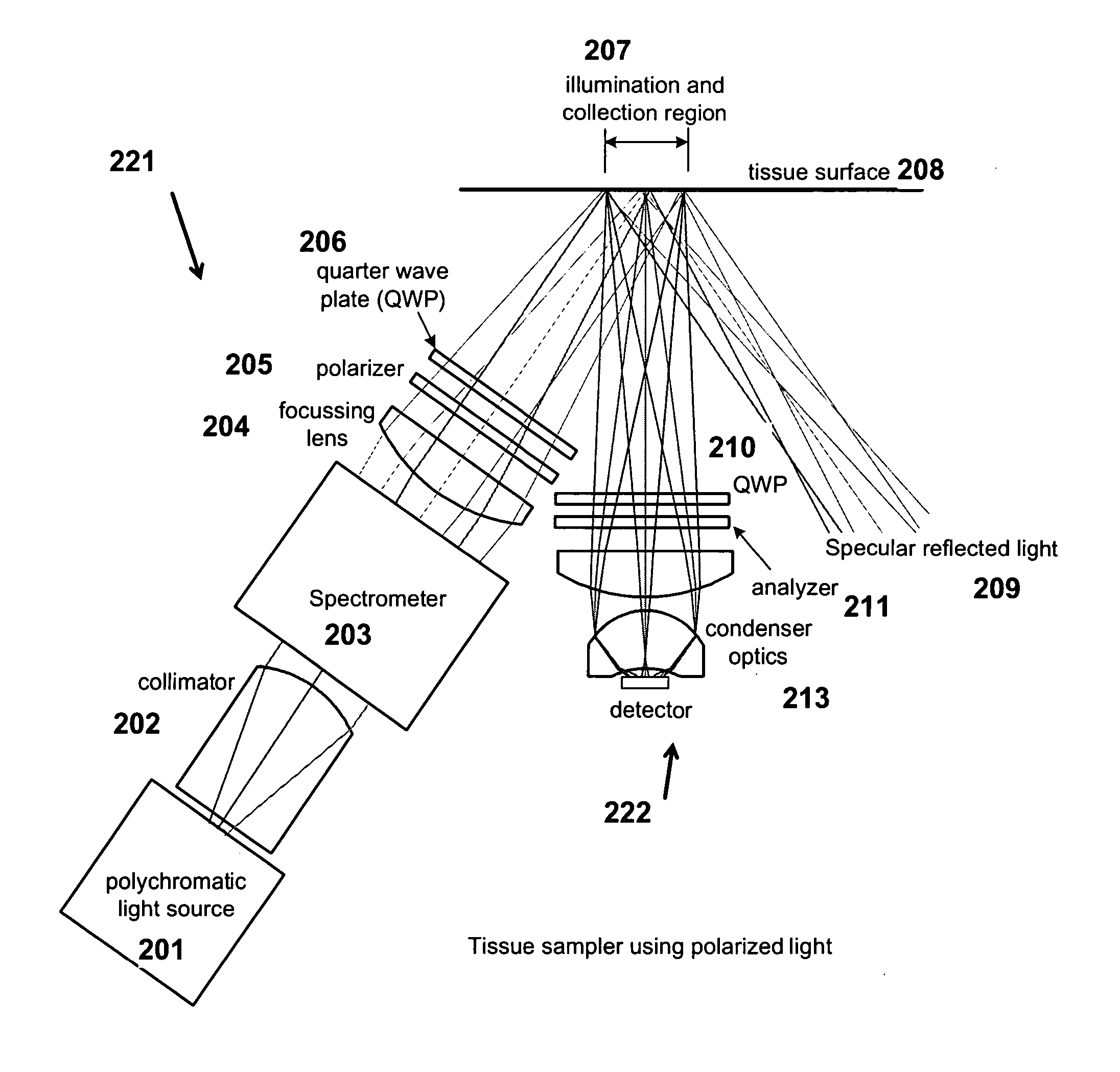
[0005] The present invention provides methods and apparatuses for accurate noninvasive determination of tissue properties. Some embodiments of the present invention comprise an optical sampler having an illumination subsystem, adapted to communicate light having a first polarization to a tissue surface; a collection subsystem, adapted to collect light having a second polarization communicated from the tissue after interaction with the tissue; wherein the first polarization is different from the second polarization. The difference in the polarizations can discourage collection of light specularly reflected from the tissue surface, and can encourage preferential collection of light that has interacted with a desired depth of penetration or path length distribution in the tissue. The different polarizations can, as examples, be linear polarizations with an angle between, or elliptical polarizations of different handedness.
[0006] A smoothing agent can be applied to the tissue surface to discourage polarization changes in specularly reflected light, enhancing the rejection of specularly reflected light by the polarization difference. The spectroscopic features of the smoothing agent can be determined in resulting spectroscopic information, and the presence, thickness, and proper application of the smoothing agent verified. The illumination system, collection system, or both, can exploit a plurality of polarization states, allowing multiple depths or path length distributions to be sampled, and allowing selection of specific depths or path length distributions for sampling. The rejection of specularly reflected light by polarization allows the sampler to be spaced from the tissue, reducing the problems attendant to contact samplers (e.g., tissue measurement trends due to pressure or heating). Separation of the sampler from the tissue enables a large area, e.g., 20 mm2, to be sampled. The illumination system and collection system can be disposed so as to communicate with different portions of the tissue surface, e.g., with portions that are separated by a fixed or variable distance.
[0007] The illumination system and collection system can be configured to optimize the sampling of the tissue, for example by changing the optical focus or the distance from the tissue surface in response to in interface quality detector (e.g., an autofocus system, or a spectroscopic quality feedback system). The portion of the tissue sampled can be identified with a tissue location system such as an imaging system that images a component of the vascular system, allowing measurements to be made at repeatable locations without mechanical constraints on the tissue.

Problems solved by technology

To date, none of these groups has demonstrated a system that generates noninvasive glucose measurements adequate to satisfy both the U.S. Food and Drug Administration (“FDA”) and the physician community.
Spectroscopic noise introduced by the tissue media is a principal reason for these failures.
Changes in the optical properties of tissue can contribute to tissue noise.
The measurement system itself can also introduce tissue noise, for example changes in the system can make the properties of the tissue appear different.
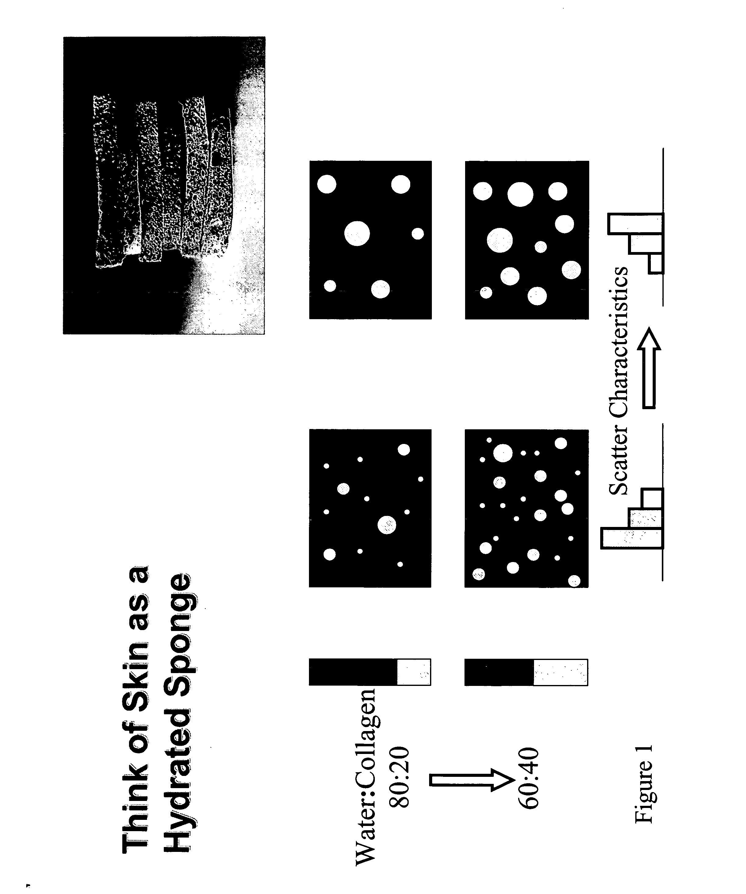
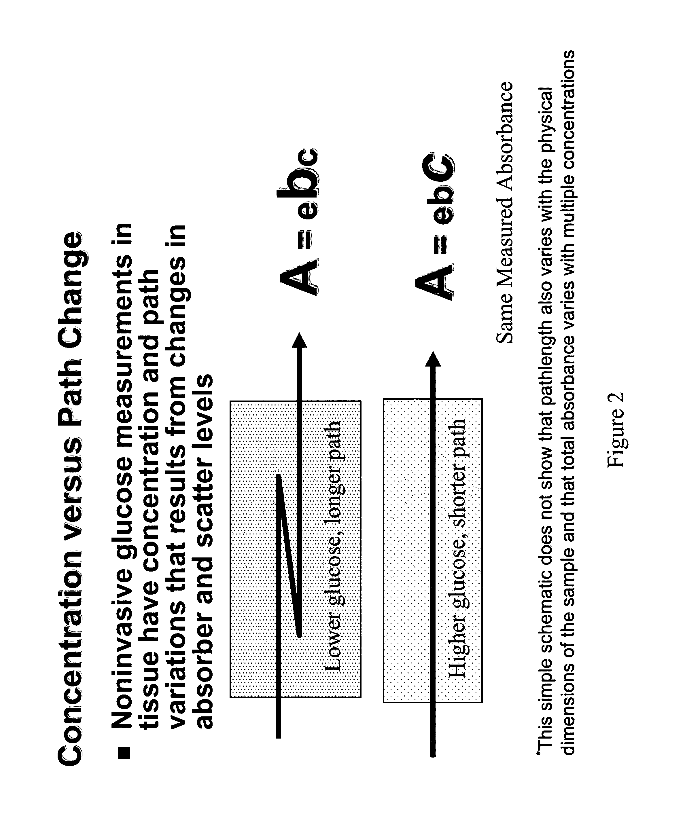
Variations in the optical properties of tissue can limit the applicability of conventional spectroscopy to noninvasive measurement.
Unfortunately, optical measurement of tissue does not match the assumptions required by Beer's law.

Method used


Image

  • Methods and apparatuses for noninvasive determinations of analytes
  • Methods and apparatuses for noninvasive determinations of analytes
  • Methods and apparatuses for noninvasive determinations of analytes

Examples


embodiments and improvements

ADDITIONAL EMBODIMENTS AND IMPROVEMENTS

[0060] A sampling system such as described in the example embodiment above can be modified for specific performance objectives by one or more of the additional embodiments and improvements described below.

[0061] Auto Focus. A motorized servo system along with a focus sensor, such as that used in autofocus cameras, can be used to maintain a precise distance between the tissue and the spectral measurement optical system during the measurement period. The tissue, the optical system, or both can be moved responsive to information from an autofocus sensor to cause a predetermined distance between the tissue and the optical system. Such an autofocus system can be especially applicable if the sampling site is the back of the hand or the area between the thumb and first finger. For example if a hand is placed on a flat surface, the auto focus mechanism could compensate for differences in hand thickness.

[0062] Tissue Scanning. The tissue can be scanne...

example embodiment

[0090]FIG. 9 is a schematic depiction of an example embodiment. This sampler eliminates the re-imaging optics of the previous sampler, bringing the light to and from the tissue by directly contacting optical fibers with the tissue. This arrangement can reduce the requirement for precision optical alignment to that required in the permanent placement of the fibers during manufacture. Physical contact can also help reduce the collection of light scattered from the tissue surface. Direct tissue contact, however, can produce tissue property changes due to interface moisture changes and compression of the underlying structure.


PUM


Abstract

The present invention provides methods and apparatuses for accurate noninvasive determination of tissue properties. Some embodiments of the present invention comprise an optical sampler having an illumination subsystem, adapted to communicate light having a first polarization to a tissue surface; a collection subsystem, adapted to collect light having a second polarization communicated from the tissue after interaction with the tissue; wherein the first polarization is different from the second polarization. The difference in the polarizations can discourage collection of light specularly reflected from the tissue surface, and can encourage preferential collection of light that has interacted with a desired depth of penetration or path length distribution in the tissue. The different polarizations can, as examples, be linear polarizations with an angle between, or elliptical polarizations of different handedness.

Description

CROSS-REFERENCE TO RELATED APPLICATIONS [0001] This application claims the benefit of U.S. provisional application “The Influence of Changing Pathlength Distributions in the Measurement of Analytes Noninvasively and Methods for Mitigation and Correction,” No. 60 / 651,679, filed Feb. 9, 2005, incorporated herein by reference.BACKGROUND OF THE INVENTION [0002] This invention relates to measurements of material properties by determination of the response of a sample to incident radiation, and more specifically to the measurement of analytes such as glucose or alcohol in human tissue. [0003] Noninvasive glucose monitoring has been a long-standing objective for many development groups. Several of these groups have sought to use near infrared spectroscopy as the measurement modality. To date, none of these groups has demonstrated a system that generates noninvasive glucose measurements adequate to satisfy both the U.S. Food and Drug Administration (“FDA”) and the physician community. Spect...

Claims


Application Information

Patent Timeline
IPC IPC(8): A61B5/00G01J4/00
CPCA61B5/14558G01N21/21G01N21/49G01N2021/4792
Inventor ROBINSON, M. RIESABBINK, RUSSELL E.JOHNSON, ROBERT D.
Owner INLIGHT SOLUTIONS
Learn More