Isolation and characterization of muscle regenerating cells
a technology characterization, which is applied in the field of isolate and characterization of muscle regenerating cells, can solve the problems of difficult identification, isolation, purification, and potential heterogeneity in the function and/or origin of sublaminar myogenic cells, and the process of bm cells to injured skeletal muscle through the generation of muscle-resident satellite cell intermediates remains controversial
- Summary
- Abstract
- Description
- Claims
- Application Information
AI Technical Summary
Benefits of technology
Problems solved by technology
Method used
Image
Examples
example 1
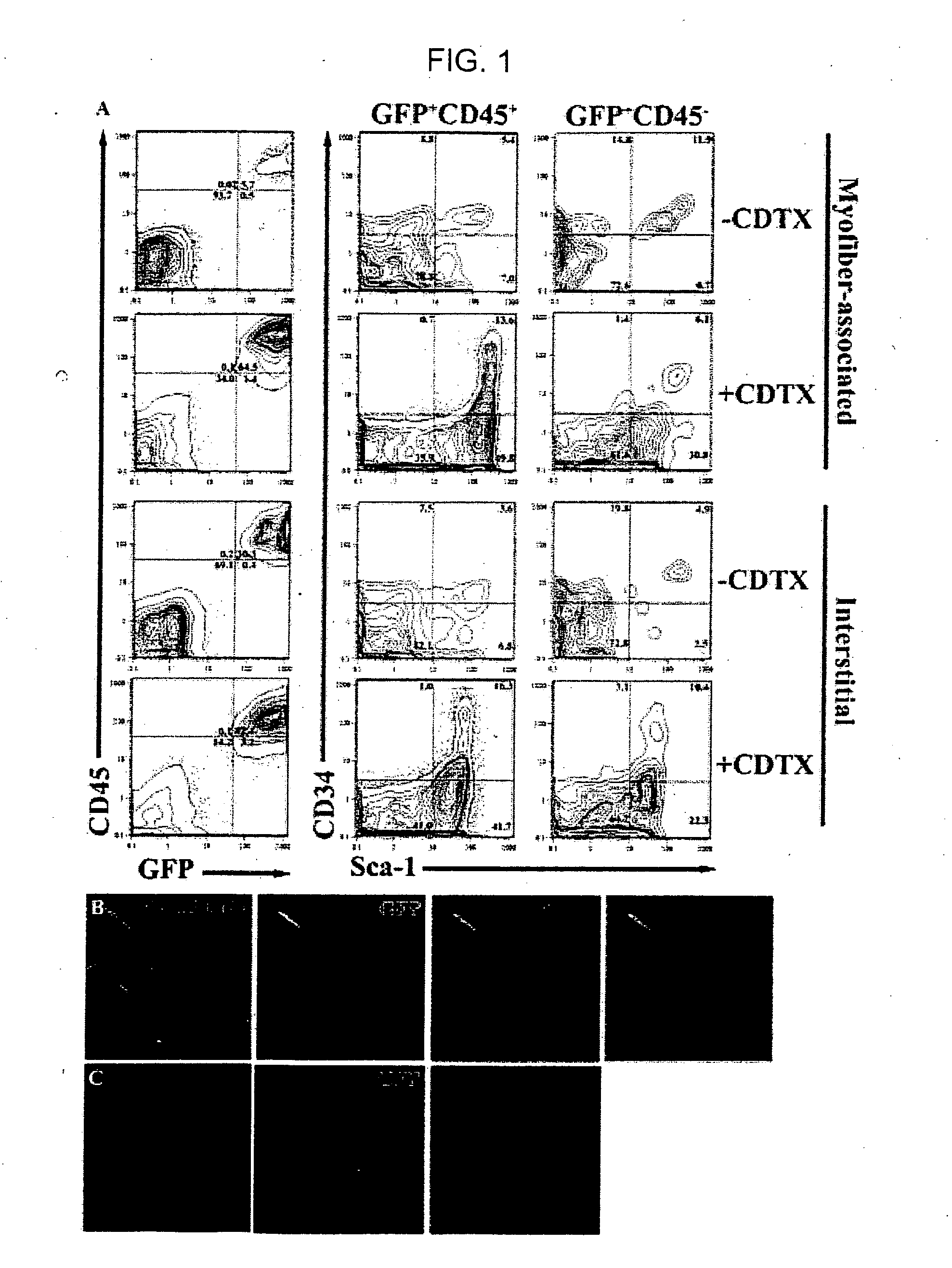
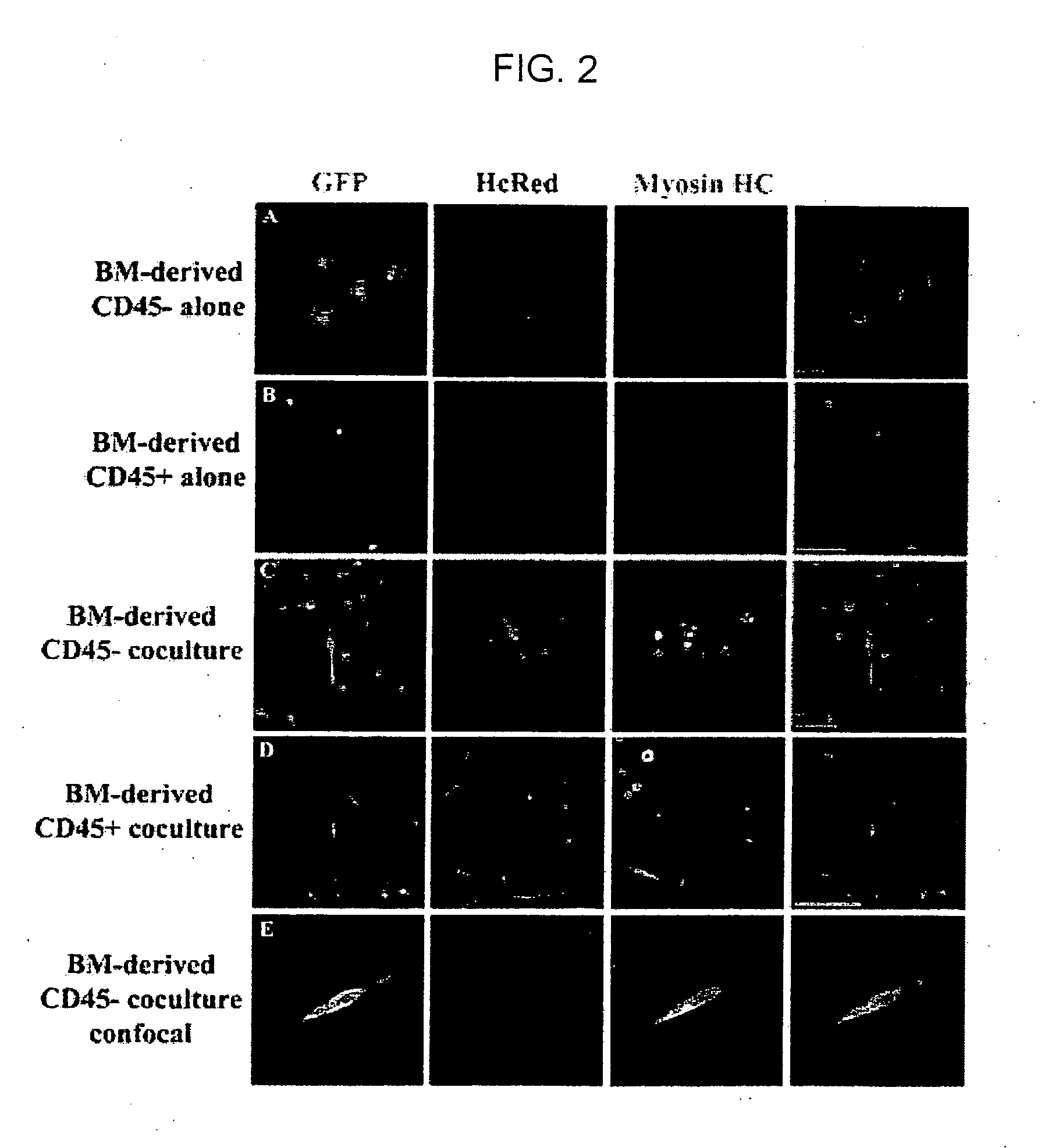
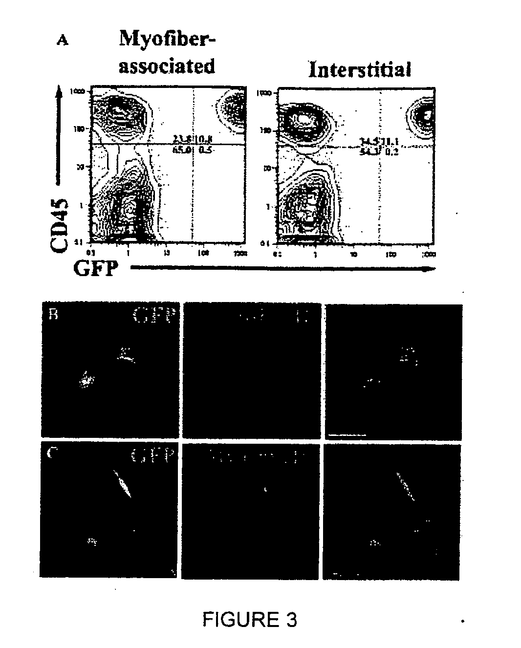
[0117] To investigate the mechanism(s) by which bone marrow (BM), hematopoietic stem cells (HSC), and circulating cells contribute to myofiber formation in injured skeletal muscle, we analyzed the myogenic potential of mononuclear cells isolated from the skeletal muscle of untransplanted mice, of mice previously transplanted with green fluorescent protein (GFP)− expressing BM or HSC, and of mice joined by parabiosis to GFP-expressing partners. GFP+ cells were found within preparations of myofiber-associated cells highly enriched for myogenic progenitor cells, and in the interstitium of injured and uninjured muscles of transplanted or parabiotic animals. Isolated muscle-engrafted cells from BM-transplanted, HSC-transplanted, or parabiotic mice generated GFP+ myofibers in vivo when injected intramuscularly into nontransgenic secondary recipients, but in contrast to endogenous myofiber-associated cells, cells engrafting muscle following transplantation or parabiosis did not express tra...
example 2
Further Characterization of Myogenic Colony Forming Cells
[0157] Myofiber associated cells were obtained by dissociation into single myofibers as described in Example 1. The cells were stained with antibodies specific for markers of interest, and tested for their ability to form myogenic colonies. The myogenic CFC were found to positively express: CD34, CXCR4, c-met (HGF-receptor), and beta1-integrin. The myogenic CFC were negative for expression of: CD45, Sca-1, Mac-1, B220, CD3, Gr-1, Thy-1, c-Kit, CD13, CD44, CD71, CD105, Flk-1, Flk-2, alpha1-integrin, and alpha6-integrin.
[0158] It was found that approximately 10% of CD45−Sca-1−CD34+ cells were myogenic CFC. In the CD45−Sca-1−Mac-1−CD34+ cell population, 18.4% give myogenic CFC. In the CD45− -Sca-1−CXCR4+ population, approximately 24% of the cells gave rise to myogenic colonies.
[0159] Cells that were sorted for the phenotype of myofiber associated, CD45−Sca-1−Mac-1− CXCR4+Beta1-integrin+ formed round myogenic colonies in 54 / 109...
PUM
| Property | Measurement | Unit |
|---|---|---|
| Fraction | aaaaa | aaaaa |
| Fraction | aaaaa | aaaaa |
| Angle | aaaaa | aaaaa |
Abstract
Description
Claims
Application Information
 - R&D
- Intellectual Property
- Life Sciences
- Materials
- Tech Scout
- Unparalleled Data Quality
- Higher Quality Content
- 60% Fewer Hallucinations
Browse by: Latest US Patents, China's latest patents, Technical Efficacy Thesaurus, Application Domain, Technology Topic, Popular Technical Reports.
© 2025 PatSnap. All rights reserved.Legal|Privacy policy|Modern Slavery Act Transparency Statement|Sitemap|About US| Contact US: help@patsnap.com


