
Materials and methods for treating vascular leakage in the eye

a technology of vascular leakage and material, which is applied in the direction of antibacterial agents, peptide/protein ingredients, dsdna viruses, etc., can solve the problems of severe vision loss and blindness, damage to and insufficient regulation of the vasculature of the ey

Inactive Publication Date: 2005-11-24
GEN VEC INC
View PDF31 Cites 8 Cited by
  • Summary
  • Abstract
  • Description
  • Claims
  • Application Information

AI Technical Summary

Problems solved by technology

Ocular-related disorders, while often not life threatening, necessitate life-style changes that jeopardize the independence of the afflicted individual.
Leading causes of severe vision loss and blindness are ocular-related disorders wherein the vasculature of the eye is damaged or insufficiently regulated.
Damage of the retina, i.e., retinal detachment, retinal tears, or retinal degeneration, is directly connected to vision loss.
Accumulation of fluid within the layers of the eye and within the vitreal cavity can cause retinal detachment, degeneration of sensory cells of the eye, increased intraocular pressure, and inflammation, all of which adversely affects vision and general health of the eye.
Prolonged periods of vascular leakage ultimately lead to thickening of the basement membrane and formation of soft and hard exudates.
Leakage of material from the vasculature into the tissues of the eye and scarring can lead to vision loss.
For many ocular-related disorders, including retinal, choroidal, and macular edema, no efficient therapeutic options currently are available.
However, laser treatment may cause permanent blind spots corresponding to the treated areas.
Laser treatment may also cause persistent or recurrent hemorrhage, increase the risk of retinal detachment, or induce neovascularization or fibrosis.
In addition, a number of patients fail to respond to laser treatment.
In most cases, all available treatment options for ocular neovascularization and uncontrolled vascular permeability have limited therapeutic effect, require repeated, costly procedures, and / or are associated with dangerous side-effects.

Method used


Image

  • Materials and methods for treating vascular leakage in the eye
  • Materials and methods for treating vascular leakage in the eye
  • Materials and methods for treating vascular leakage in the eye

Examples


example 1

[0114] This example illustrates a preferred method of obtaining expression of a factor comprising both anti-angiogenic and neurotrophic activity from an adenoviral vector in in vivo retina.

[0115] An adenoviral vector deficient in one or more essential gene functions of the E1, E3, and E4 regions of the adenoviral genome and comprising a PEDF gene (Ad.PEDF) is preferably constructed as set forth in WO 99 / 15686 (McVey et al.). However, the method of the invention is not dependent on the method of vector construction employed and previously described methods of vector construction are also suitable.

[0116] Several in vivo models of ocular neovascularization are available. Neovascularization of the retina is obtained in, for example, neonatal animals, i.e., neonatal mice, by exposing the mice to hypoxic conditions shortly after birth. Several days later, the neonatal mice are exposed to standard atmospheric conditions, resulting in ischemia-induced neovascularization of the retina.

[01...

example 2

[0119] This example demonstrates a preferred method of obtaining expression of a factor comprising both anti-angiogenic and neurotrophic activity from an adenoviral vector in in vivo choroid. The following example further provides methods for determining the effect of PEDF on neovascularization.

[0120] An adenoviral vector deficient in one or more essential gene functions of the E1, E3, and E4 regions of the adenoviral genome and comprising a PEDF gene (Ad.PEDF) is constructed as set forth in WO 99 / 15686 (McVey et al.).

[0121] An in vivo model of choroidal neovascularization can be obtained by detaching the retina of an eye of, for example, a mouse or rabbit, and debriding the pigmented epithelia. Choriocapillary regeneration is monitored in both treated and untreated eyes. Ad.PEDF is administered prior to perturbing the retinal pigment epithelial (RPE) to determine the effect of the inventive method in preventing choroidal neovascularization. Of course, Ad.PEDF is administered afte...

example 3

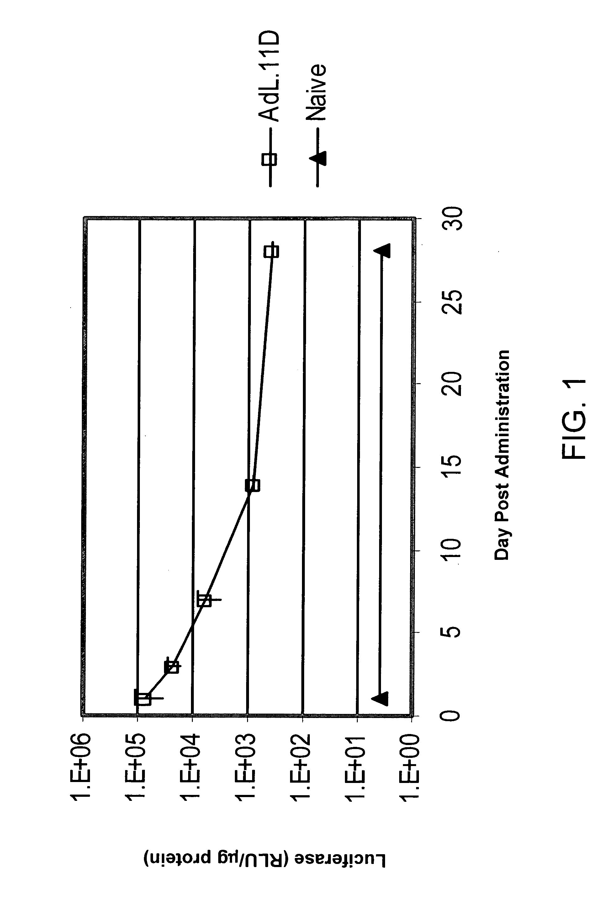
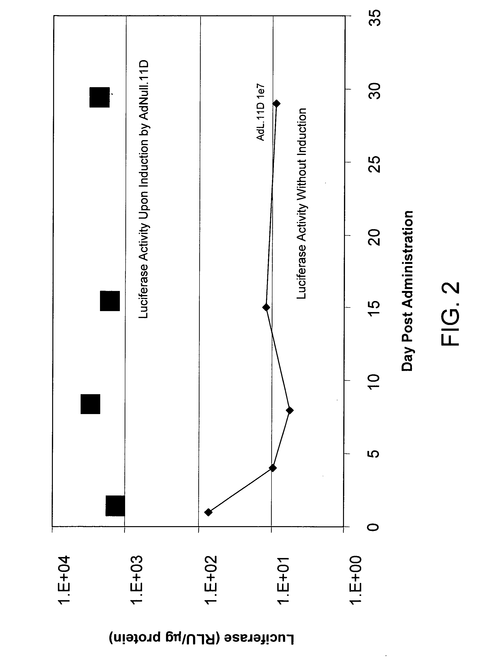
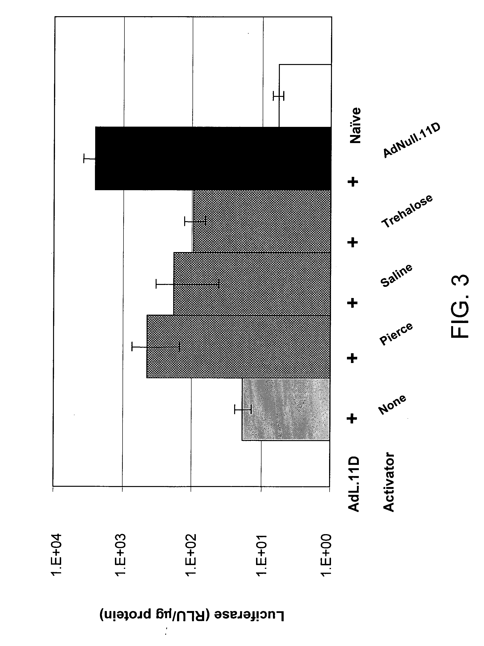
[0123] This example demonstrates the utility of adenoviral vectors to deliver multiple doses of an exogenous nucleic acid to the eye.

[0124] Adenoviral vectors comprising the luciferase gene (Ad.L) or control adenoviral vectors comprising no transgene (Ad.null) were injected into the intravitreal space of C57BL6 mouse eyes (Day 0). One day following injection of the adenoviral vectors (Day 1), eyes infected with Ad.L were enucleated and frozen (1st administration). The eyes infected with Ad.null were divided into three groups. In Group I, Ad.L vectors were injected into the intravitreal space of the eye at Day 14 (fourteen days following the initial dose of Ad.null). Group I eyes were enucleated and frozen the day following the second administration of adenoviral vectors (Day 15, 2nd administration). Group II eyes were injected intravitreally with Ad.null at Day 14, and injected intravitreally with Ad.L vectors four weeks following the initial injection with Ad.null (Day 28, 3rd adm...


PUM

PropertyMeasurementUnit
pHaaaaaaaaaa
ionic strengthaaaaaaaaaa

Abstract

The invention is directed to a method of prophylactically or therapeutically treating an animal for vascular leakage in an eye. The method comprises administering to an animal in need thereof an expression vector comprising a nucleic acid sequence encoding pigment epithelium-derived factor (PEDF) such that vascular leakage in the eye of the animal is treated prophylactically or therapeutically. The invention also provides a method of prophylactically or therapeutically treating an animal for vascular leakage in an eye. The method comprises periocularly administering to an animal in need thereof PEDF such that vascular leakage of the animal is treated prophylactically or therapeutically. The invention also provides a method of prophylactically or therapeutically treating an animal for non-diabetic vascular leakage in an eye. The method comprises administering to an animal in need thereof PEDF such that non-diabetic vascular leakage in the eye is treated prophylactically or therapeutically.

Description

CROSS-REFERENCE TO RELATED PATENT APPLICATIONS [0001] This patent application claims the benefit of U.S. Provisional Patent Application No. 60 / 552,768, filed Mar.12, 2004.FIELD OF THE INVENTION [0002] The invention relates to a method of prophylactically or therapeutically treating an animal for vascular leakage in the eye. BACKGROUND OF THE INVENTION [0003] An overwhelming majority of the world's population will experience some degree of vision loss in their lifetime. Vision loss affects virtually all people regardless of age, race, economic or social status, or geographical location. Ocular-related disorders, while often not life threatening, necessitate life-style changes that jeopardize the independence of the afflicted individual. Vision impairment can result from most all ocular disorders, including diabetic retinopathies, proliferative retinopathies, retinal detachment, toxic retinopathies, retinal vascular diseases, retinal degenerations, vascular anomalies, age-related macu...

Claims


Application Information

Patent Timeline
Patent Type & Authority Applications(United States)
IPC IPC(8): A61K38/17A61K38/57A61K48/00C12N15/861
CPCA61K9/0051A61K38/1709A61K38/57C12N2710/10343A61K48/005C12N15/86A61K48/00A61P15/08A61P27/02A61P27/12A61P29/00A61P31/04A61P31/18A61P35/00A61P43/00A61P9/10
Inventor WEI, LISARASMUSSEN, HENRIK
Owner GEN VEC INC
Learn More