
Shaping suture for treating congestive heart failure

a congestive heart failure and suture technology, applied in the field of congestive heart failure treatment devices and methods, can solve the problems of congestive heart failure, poor overall treatment effect of all available therapies, and inability to improve the function of weakened heart muscle, etc., to achieve the effect of improving the overall treatment of an ischemic heart and enhancing the overall treatmen

Inactive Publication Date: 2005-05-26
CHF TECH A CALIFORNIA CORP
View PDF20 Cites 86 Cited by
  • Summary
  • Abstract
  • Description
  • Claims
  • Application Information

AI Technical Summary

Benefits of technology

[0019] The present invention endeavors to address those deficiencies as well as improve and enhance the overall treatment of an ischemic heart. In one embodiment, the present invention comprises a kit comprising multiple components, as well as a method for providing and / or utilizing one or more of the components, for treating ischemic congestive heart failure. This kit and the method of providing and using the kit can aid a practitioner in excluding and reinforcing the akinetic portion of a heart chamber, a procedure sometimes referred to as Surgical Ventricular Restoration (SVR), without creating a heart chamber that is too small or too spherical.
[0020] In one embodiment of the invention, the kit comprises a device for sizing and shaping a deficient ventricle. One benefit of certain embodiments of the shaping device of this invention, discussed in more detail below, is that they do not require inflation. Unlike inflatable shaping devices, there is no risk of puncturing and deflating this device during the procedure. The shaping device of this invention can also be compliant unlike inflatable devices that must be inflated to a point at which they become non-compliant. The kit may also comprise a device for deploying and removing the shaping device.
[0022] The method of the present invention may comprise steps that utilize one or more of the following components: a shaper, a patch, and a shaping suture. These components can aid in creating a heart chamber of the appropriate shape and size. The present method may comprise the step of determining an appropriate size and shape for a heart chamber based on the needs of the patient. A practitioner may use any combination of methods for determining the appropriate heart chamber size and shape for the patient. Some potential methods include but are not limited to magnetic resonance imaging (MRI), PET Scan, Echo, ultrasound, end diastolic volume, and / or body surface measurement. Images of the heart chamber may be provided to a computer. Computer software, databases, or computer networks may aid in determining an appropriate size and shape as well. The practitioner may then choose a correctly sized shaping device, suture, and / or patch for the patient.
[0025] The method may further comprise the step of weaving a purse-string stitch around the border between the akinetic or dyskinetic tissue and the healthier tissue. In weaving the purse-string stitch, the practitioner preferably excludes the akinetic tissue and reshapes the heart chamber by pulling the chamber wall together around the shaping device. The practitioner may use more than one row of purse-string stitches as needed. Preferably, the practitioner can use a shaping suture to weave the purse-string stitch, which can aid in forming an oblong rather than circular shape when pulling tissue together with the purse-string stitch.

Problems solved by technology

Unfortunately, the collective results of all available therapies in the treatment of congestive heart failure are disappointing.
Pharmacology and electrical resynchronization have improved the symptoms in many cases, but direct approaches to improving the function of the weakened heart muscle, the common thread in all cases, are few.
One common cause of congestive heart failure is a previous heart attack causing “ischemia,” or lack of oxygen to the heart tissue.
When treated, the patient is stabilized, often receiving a balloon catheter dilitation, intra-coronary stent or bypass graft, and has an initially unremarkable recovery.
Drugs such as ARBs (angiotensin receptor blockers) and ACE (angiotensin converting enzyme) inhibitors have been shown to retard the progress of this disintegration of cardiac function, but the end result is delay, not cure.
In diseased hearts, the compensation is not sufficient and the ventricular dilation and muscle hypertrophy progress to a point where efficiency of heart function begins to fall.
Further attempts by the heart to compensate may accelerate this reduction in efficiency.
First, it is difficult for surgeons using the procedure to reshape the ventricle to its naturally elongated shape.
Without the elongated, conical shape normally associated with a healthy heart, the ventricle cannot perform the twisting motion at the apex that can account for a large percentage of the pumping action.
A more spherical ventricle must rely almost entirely on lateral squeezing action, which is very inefficient.
Some surgeons inaccurately estimate the appropriate ventricle size resulting in a ventricle that is too small, which may leave the patient clinically worse than before the procedure.
Other surgeons fail to account for the desired shape of the ventricle and do nothing to try to achieve a less spherical shape.
The use of a balloon, however, has not adequately solved the problem.
First, it does not aid the practitioner in achieving a less spherically shaped ventricle.
Second, the practitioner must still estimate the appropriate size for the ventricle in deciding how much to fill and expand the balloon.
Finally, the balloon has the added disadvantage that a needle or any other sharp object used during the procedure may rupture the balloon and render it useless for the remainder of the procedure.

Method used


Image

  • Shaping suture for treating congestive heart failure
  • Shaping suture for treating congestive heart failure
  • Shaping suture for treating congestive heart failure

Examples


Embodiment Construction

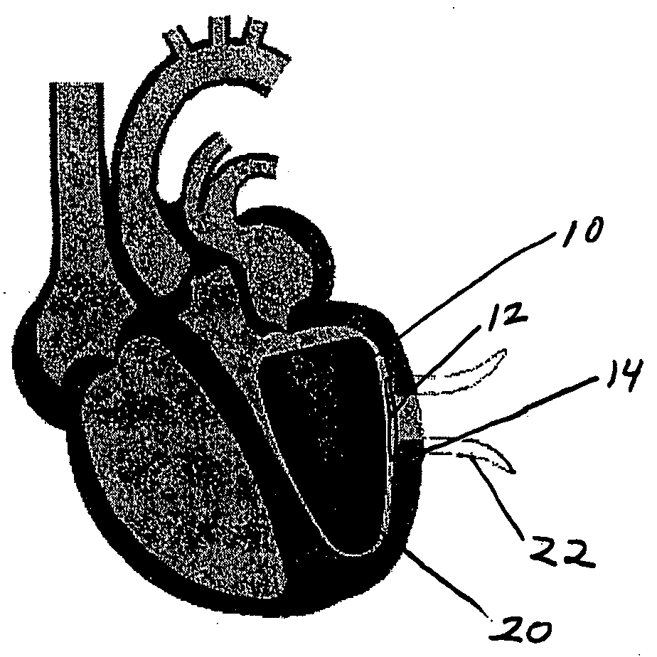
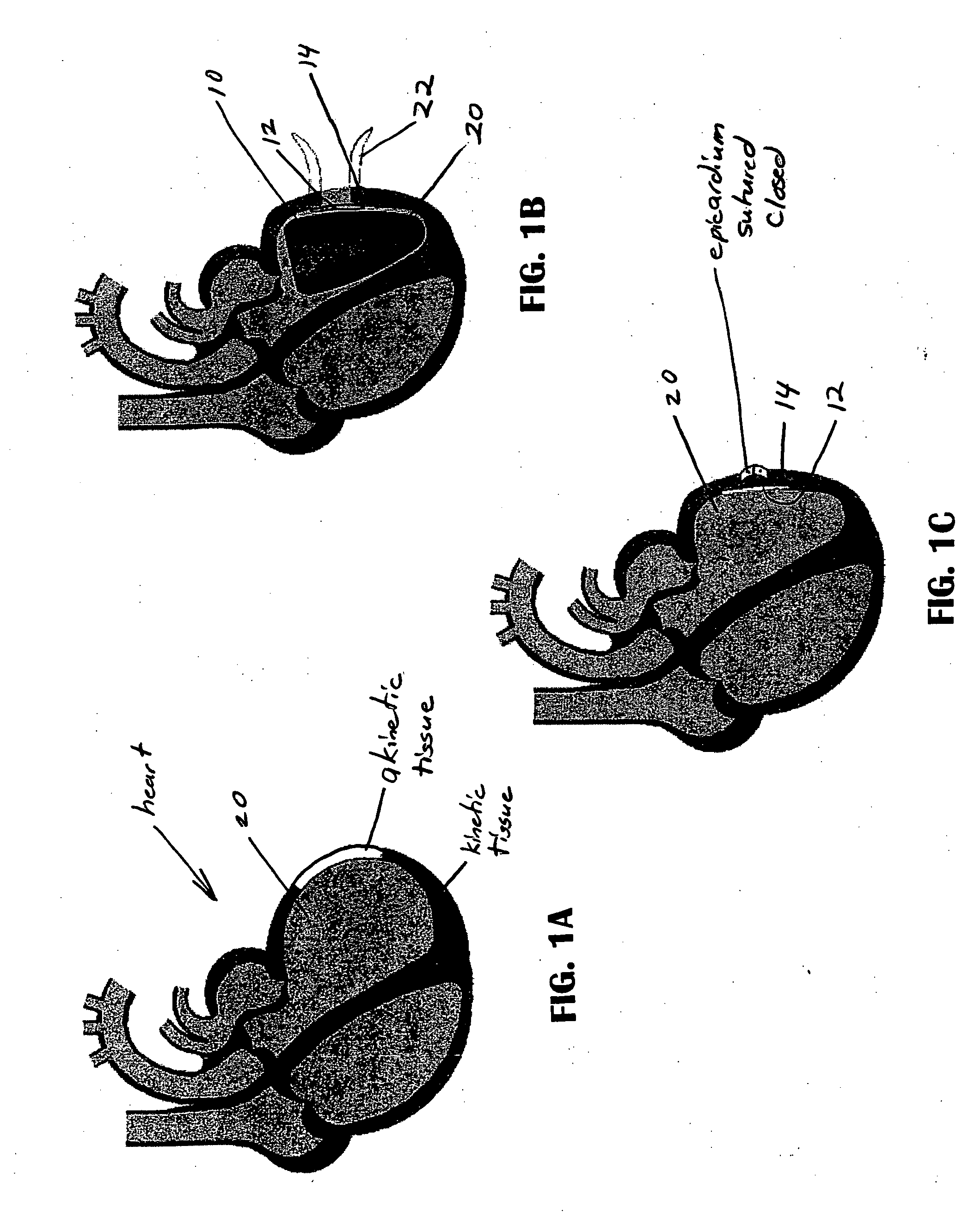
[0047] The present invention allows more exact execution of a procedure or process whose purpose is to create a more optimum size and shape of an organ, such as the heart or more particularly a heart chamber, or another structure undergoing reconstruction. For example, one such procedure is known as Surgical Ventricular Restoration (SVR). Referring to FIGS. 1A-1C, the invention comprises a kit and a method of using the kit. The kit may include a shaping device 10, described in more detail below in association with FIGS. 3-7. In addition, the kit may include a patch 12, described in more detail below in association with FIGS. 8-11. The kit may further include a shaping suture 14, described in more detail below in association with FIGS. 12-15. In some embodiments, the kit may also include an integrated shaping device and patch, described in more detail below in association with FIG. 16. Some embodiments of the kit may further include a patch sizing template, described below in more de...


PUM


Abstract

A kit and method are described for treating congestive heart failure. The kit may comprise multiple components including a shaping device, deployment tool, patch, and suture. The method may utilize one or more of these components.

Description

RELATED APPLICATIONS [0001] This application claims priority from the following U.S. Provisional Patent Applications each of which is incorporated herein in its entirety by reference: Ser. No. 60 / 466,653, filed Apr. 29, 2003 and titled Ventricular Restoration; Ser. No. 60 / 485,568, filed Jul. 7, 2003 and titled Systems, Devices and Methods of Use for Treating Congestive Heart Failure (CHF); Ser. No. 60 / 488,292, filed Jul. 18, 2003 and titled Ventricular Sizing & Shaping Device and Method; Ser. No. 60 / 499,946, filed Sep. 2, 2003 and titled System and Method of Use to Employ Imaging Technology for Diagnosis, Measurement, Standardization, and Follow-up of Disease Processes and Determine Optimal Treatment; Ser. No. 60 / 500,762, filed Sep. 4, 2003 and titled Shaping Suture Device and Method of Use; Ser. No. 60 / 512,293, filed Oct. 17, 2003 and titled Less Invasive CHF Treatment—Reshaping the Heart; Ser. No. 60 / 518,270, filed Nov. 5, 2003 and titled Methods and Devices for Tracking Acute Myo...

Claims


Application Information

Patent Timeline
Patent Type & Authority Applications(United States)
IPC IPC(8): A61B17/00
CPCA61B17/0057A61B2017/00663A61B2017/00659
Inventor HOUSER, RUSSELL A.BERTOLERO, ARTHUR A.ANNEST, LONHARE, BILLIBRAHIM, TAMERGEYSTER, STEVESMITH, WENDEL
Owner CHF TECH A CALIFORNIA CORP
Learn More