
Cervical canal dilator

a cervical canal and dilation device technology, applied in the field of cervical dilation devices, can solve the problems of not keeping well-defined shapes, unable to be used to exert high pressure in medical applications, and limited gutnicks

Inactive Publication Date: 2005-03-10
SIEMENS MEDICAL SOLUTIONS USA INC
View PDF4 Cites 56 Cited by
  • Summary
  • Abstract
  • Description
  • Claims
  • Application Information

AI Technical Summary

Benefits of technology

[0030] A control system is connected with the at least two lumens and includes means for a f...

Problems solved by technology

They do not retain well-defined shapes, though, and cannot be used to exert high pressure in medical applications.
Gutnick, however, is limited by the combination of the structural material of the dilating member being elastic and the inflation process of supplying a controlled volume of liquid to the elastic dilating member to produce an inflated diameter of the elastic member which is stated in one example as “about somewhat greater than 10 mm and preferably. expand up to about 15 mm.” Thus, the structure of the Gutnick elastic dilating member and the inflation process thereof is limited in its ability to accurately produce a specific or controlled desirable maximum inflation diameter.
The Gutnick method to determine the diameter of inflation relative to a given volume is not directly measured and thus is highly subjective, vulnerable to varying lengths of conduits and fluid losses and is therefore also vulnerable to being overly expanded and damaging the cervix.
This can result in a partial or an uneven dilation of the cervix because the combination of the length of the second inflatable member relative to its placement in the cervix can be too short to adequately treat all cervixes.
Finally, the disc member limits the visualization of the positioning of the dilator into the cervical canal adding further risk of harm to the patient.
Levine is limited by its inability to dilate the cervical opening beyond the diameter of the shaft.
In addition, the limited range of the angle of inclination of the distal end between 15 and 25 degrees also inhibits the flexibility in which Levine can be applied due to natural variations in the orientation of the cervix to the axis defined by the vagina.
In addition, the balloons or first inflatable member and second expandable member lack the ability to provide an indication as to how much compressive pressure they are applying against the cervix while securing the shaft.
The ability of the metal rod to penetrate beyond the tip of the shaft and damage the uterus also presents a potential safety hazard.
The ability of the ripening device of Cowan to provide uniform pressure along the length of the cervix in all situations is questionable.
The application of this shape of device may unevenly dilate the cervix by over dilating the edges and under dilating the central portion.
Under dilating can complicate the passage of instruments.
Uneven dilation can cause discomfort to the patient and damage to the cervix.
Further, the shape of the balloon inhibits the ability of the physician to monitor the amount of dilation being achieved by the device.
Overly dilating the cervix can cause damage to the cervix.
Despite historical uses of inelastic balloons in medicine, there have been limitations in the use of these balloons for dilation of the cervical canal.
This challenge arises due to the lack of visualization of the cervix to assist the user in placing the catheter, whereas in vascular applications such as angioplasty the catheter placement can be visualized using fluoroscopy.
For this reason, a single balloon for dilation, such as those used in angioplasty, are ineffective, resulting in a potential to either insert the catheter too far, causing damage to the uterus, or fail to dilate the full length of the cervix, if the catheter is not placed far enough into the cervix.
Prior attempts to overcome this lack of visualization through the use of multiple balloons or unique balloon shapes have been limited by the unique problems with cervical dilation occurring because there is more resistance to dilation of the internal os (portion of the cervical canal adjacent to the uterus) which is furthest from the operator, than the portion of the cervix closer to the vagina.
The unequal resistance tends to push a balloon out toward the vagina, so patients may not have their inner os properly dilated.
The inelastic ellipsoidal balloon is intended to act as an anchor and to dilate the internal os, but use of an ellipsoidal balloon to inflate the internal os may result in an overdilation of the internal os, risking damage to the cervix including an incompetent cervix (a cervix unable to remain closed for a fetus causing miscarriage).
If the ellipsoidal balloon is used as an anchor for positioning, use of this design results in under-inflation of the inner os because inelastic balloons have a taper to allow the folding of the balloon when deflated.
The taper of the balloons results in a set gap between the two balloons at the point of the inner os, resulting in an under-inflated portion of the cervix.
Another problem encountered with the use of inelastic balloons is that the balloons tend to be fragile.
These instruments can damage the balloons leading to balloon rupture.
One reason for this is that the primary procedure is angioplasty, where the use of air would risk the release of a potentially life-threatening air embolus if the balloon ruptured.

Method used


Image

  • Cervical canal dilator
  • Cervical canal dilator
  • Cervical canal dilator

Examples


Embodiment Construction

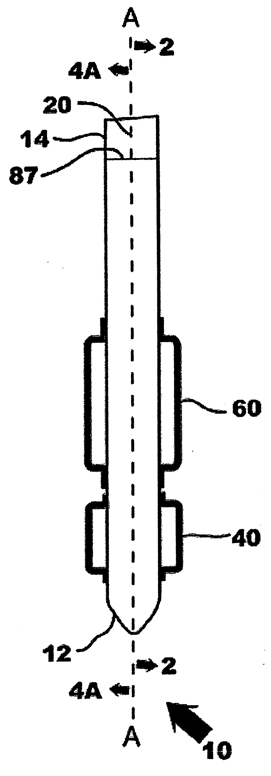
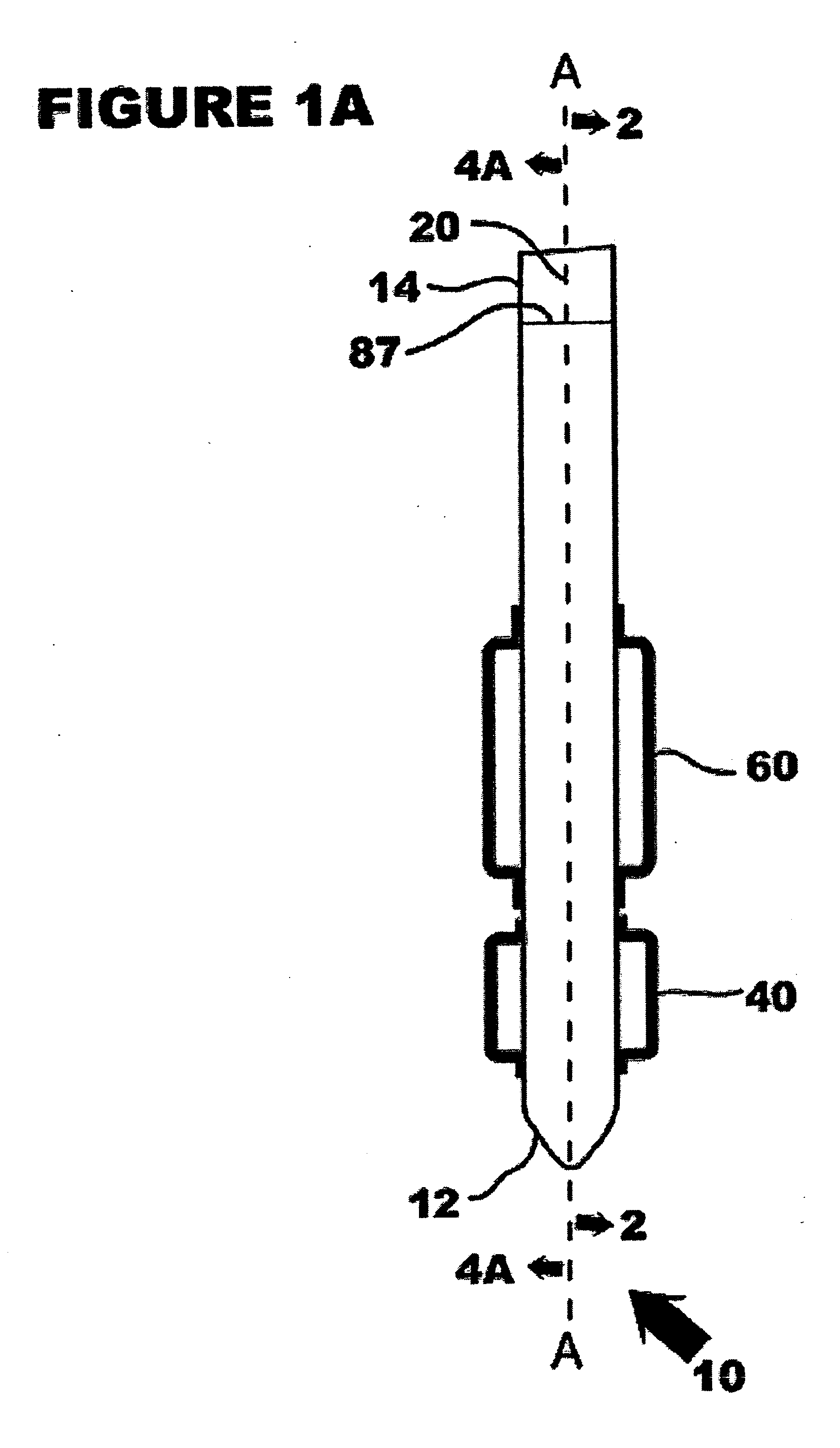
[0042] Referring now in specific detail to the drawings in which like referenced numerals identify similar or identical elements throughout the several views, and initially to FIG. 1A, a novel cervical canal dilator assembly 10 is shown having a shaft 20, a first inflatable member 40, a second inflatable member 60 and a control system 90 (see FIG. 3). Cervical canal dilator assembly 10, hereinafter referred to as “dilator 10” has a distal end 12 and a proximal end 14 defining a longitudinal axis-A.

[0043] In FIG. 1B, the novel cervical canal dilator assembly 10 is shown having a sheath 80. Sheath 80 includes markings 88 for correlating the position of the first inflatable member relative to the proximal end of the sheath. Shaft 20 includes markings 87 for correlating the position of sheath 80 relative to the inflatable members. Dilator 10 is adapted for use by a physician and is configured as a readily useable disposable device having a reduced cross-sectional dimension of less than...


PUM


Abstract

A cervical canal dilating assembly and method of use are shown. The dilator assembly includes a plastic shaft, a first inflatable member, and a second inflatable member. The shaft can range from being rigid to being highly flexible. The second inflatable member is fabricated of a non-elastic material and is configured to have a maximum inflatable diameter. The second inflatable member is configured to have a predetermined maximum inflatable diameter ranging from 4 to 20 mm. The dilating assembly can also be at least partially covered by a sheath. A control system includes means for measuring pressure configured for at least monitoring the pressure of the second inflatable member. A wire can be used in selected configurations to stiffen and shape the shaft. In operation, the initial penetration of the dilating assembly into the uterus uses a wire for increased stiffness. The dilating assembly is then forwarded through the remainder of the cervical canal. The first inflatable member is expanded in the uterus after being uncovered by the sheath. The second inflatable member is positioned in the cervical canal and gradually inflated to a predetermined maximum diameter.

Description

FIELD OF THE INVENTION [0001] The present disclosure relates to devices for cervical dilation. More particularly, the present disclosure relates to devices for cervical dilation using inflatable members. BACKGROUND OF THE INVENTION [0002] Dilators for body passageways are well known and have many functions in medicine. The use of elastic balloons, made of materials such as latex rubber or silicone, is well established in medicine, typically for applications where low pressures are needed for fixation and occlusion. Elastic balloons are inflated by volume and can typically stretch 100--600%. They do not retain well-defined shapes, though, and cannot be used to exert high pressure in medical applications. When the pressure is released, elastic balloons recover close to their original size and shape. An example of an elastic balloon is seen with Foley catheters that are passed through the urethra into the bladder; an elastic balloon located near the distal tip is inflated to occlude th...

Claims


Application Information

Patent Timeline
IPC IPC(8): A61BA61F2/958A61M29/00A61M29/02A61M31/00
CPCA61M25/1011A61M25/1018A61M2025/0063A61M31/00A61M29/02A61M25/10181A61M25/10184
Inventor FOLTZ, JONATHANREED, DAVIDOLIVE, DAVID
Owner SIEMENS MEDICAL SOLUTIONS USA INC
Learn More