
Method and apparatus for non-invasively evaluating endothelial activity in a patient

a non-invasive, patient technology, applied in the field of non-invasive evaluation of endothelial activity in patients, can solve the problems of high cost, high labor intensity, and high labor intensity, and achieve the effect of improving the test results

Inactive Publication Date: 2004-05-13
ITAMAR MEDICAL LTD
View PDF7 Cites 51 Cited by
  • Summary
  • Abstract
  • Description
  • Claims
  • Application Information

AI Technical Summary

Benefits of technology

[0036] FIG. 12 diagrammatically illustrates one manner of improving the performance of the described non-invasive technique in evaluating endothelial activity, in which a reference digit probe is used on a finger of a non-occluded arm of the patient;
[0037] FIG. 13 illustrates another manner of improving the test results by including the reference digit-probe on a second finger of the same hand of the patient;

Problems solved by technology

No currently available test of endothelial function is suitable for wide clinical application for determining the presence of ED, which is now well accepted as an important factor in the pathogenesis of atherosclerotic cardiovascular disease.
The tests currently available are either highly invasive (i.e., measuring the hemodynamic changes caused by intracoronary instillation of vasoactive drugs), or require the use of technically difficult vascular imaging studies, expensive apparatus, and highly skilled staff.
For example, it requires expensive apparatus and specialized personnel, and it suffers from a lack of accuracy and poor inter and intra-observer reproducibility.
The method is also very uncomfortable to the subject since the pressure cuff is very tight around the subject's arm and blood flow must be stopped for a relatively long time, e.g., 5 minutes.

Method used


Image

  • Method and apparatus for non-invasively evaluating endothelial activity in a patient
  • Method and apparatus for non-invasively evaluating endothelial activity in a patient
  • Method and apparatus for non-invasively evaluating endothelial activity in a patient

Examples


Embodiment Construction

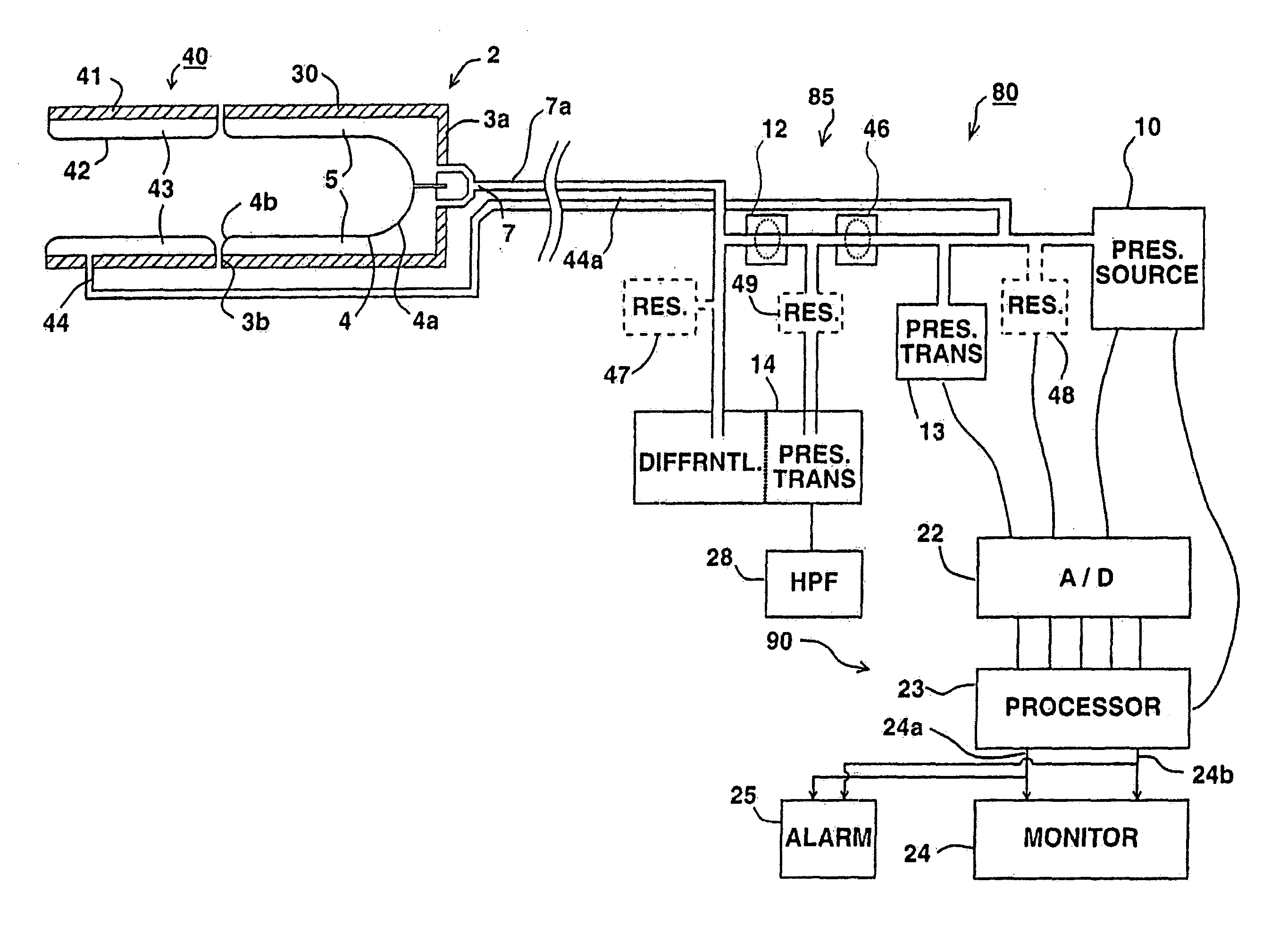
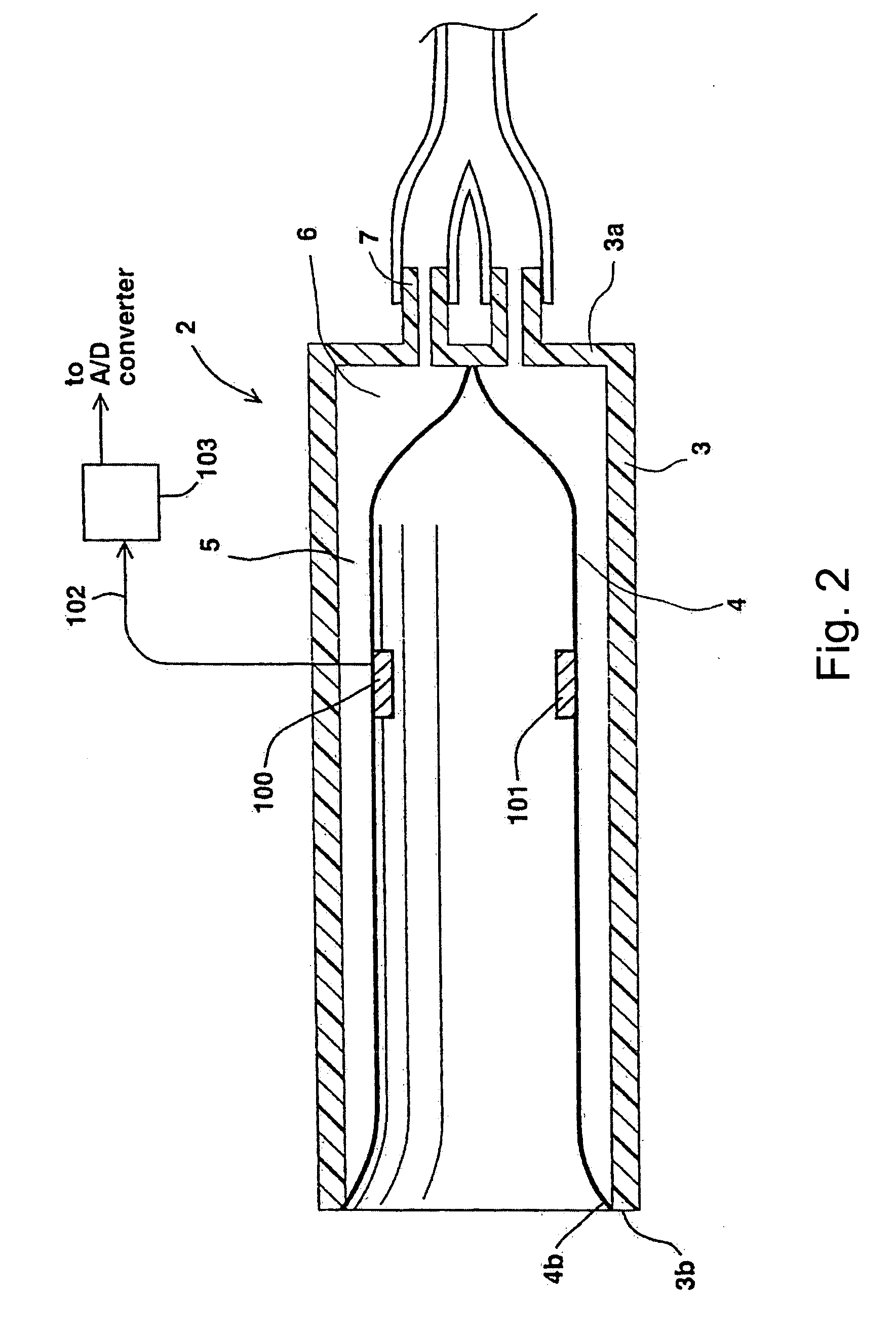
[0044] As briefly described above, and as more particularly described below, the present invention provides a method for non-invasively evaluating endothelial activity in a patient, particularly for indicating the presence of an endothelial dysfunction condition. This is done non-invasively by: applying an occluding pressure to a predetermined part of an arm or leg of the patient to occlude an arterial blood flow therein; maintaining the occluding pressure for a predetermined time period; removing the occluding pressure after the elapse of the predetermined time period to restore arterial blood flow; monitoring a digit of the arm or leg for changes in the peripheral arterial tone therein before, during, and after the application of the occluding pressure to the arm or leg of the patient; and utilizing any detected changes in the peripheral arterial tone for evaluating endothelial activity in the patient, and particularly for indicating the presence of an endothelial dysfunction cond...


PUM


Abstract

A method and apparatus for non-invasively evaluating endothelial activity in a patient, particularly for indicating the presence of an endothelial dysfunction condition, by applying an occluding pressure to a predetermined part of an arm or leg of the patient to occlude arterial blood flow therein; maintaining the occluding pressure for a predetermined time period; removing the occluding pressure after the elapse of the predetermined time period to restore arterial blood flow; monitoring a digit of the arm or leg by a digit-probe for changes in the peripheral arterial tone therein before and after the application of the occluding pressure to the arm or leg of the patient; and utilizing any detected changes in the peripheral arterial tone for evaluating endothelial activity in the patient. Particularly important advantages are provided when the occluding cuff is applied to the digit of the patient receiving the monitoring digit-probe, on the proximal side thereof with respect to the patient's heart, and also when a reference digit-probe is applied to another digit, not influenced by the occluding pressure, to compensate for shifts inherent to vascular beds.

Description

[0001] The present invention relates to the method and apparatus of PCT Application No. PCT / IL97 / 00249, International Publication No. WO 98 / 04182, published 5 Feb. 1998, and PCT Application No. PCT / IL00 / 00307, International Publication No. WO 00 / 74551, published 14 Dec. 2000, which applications are hereby incorporated by reference as if fully set forth herein.[0002] The above PCT applications relate to the non-invasive detection and monitoring of various physiological states or medical conditions of a patient by using a digit-probe for monitoring the peripheral arterial tone (PAT) of the patient. The present invention relates to non-invasively evaluating endothelial activity in a patient, particularly for indicating the presence of an endothelial dysfunction condition, preferably by using the digit-probe of those applications.[0003] The active control of vascular smooth muscle (VSM) tone in the arteries of the finger (or toe) is by way of alpha-adrenergic sympathetic nervous control...

Claims


Application Information

Patent Timeline
Patent Type & Authority Applications(United States)
IPC IPC(8): A61B5/022A61B5/0245A61B5/026
CPCA61B5/02007A61B5/02241A61B5/026A61B5/6838A61B5/6826A61B5/6828A61B5/6824
Inventor SCHNALL, ROBERT PSHEFFY, JACOBLAVIE, PERETZ
Owner ITAMAR MEDICAL LTD
Learn More