Imaging methods and apparatus particularly useful for two and three-dimensional angiography

Inactive Publication Date: 2003-03-13
MEDIMAG C V I
View PDF1 Cites 20 Cited by
  • Summary
  • Abstract
  • Description
  • Claims
  • Application Information

AI Technical Summary

Benefits of technology

[0010] An object of the present invention is to provide an improved method and apparatus for producing two-dimensional or three-dimensional angiographic images to better enable diagnosis or treatment of a disease in the human body. A further object of the invention is to provide a method and apparatus for enhancing the CNR of a DSA image.

Problems solved by technology

However, using a respiration sensor as the reference is not precise because of the difficulty in sharply defining a particular point of the respiration curve for this purpose.

Method used


Image

  • Imaging methods and apparatus particularly useful for two and three-dimensional angiography
  • Imaging methods and apparatus particularly useful for two and three-dimensional angiography
  • Imaging methods and apparatus particularly useful for two and three-dimensional angiography

Examples


Embodiment Construction

[0038] The Embodiment of FIGS. 1-6

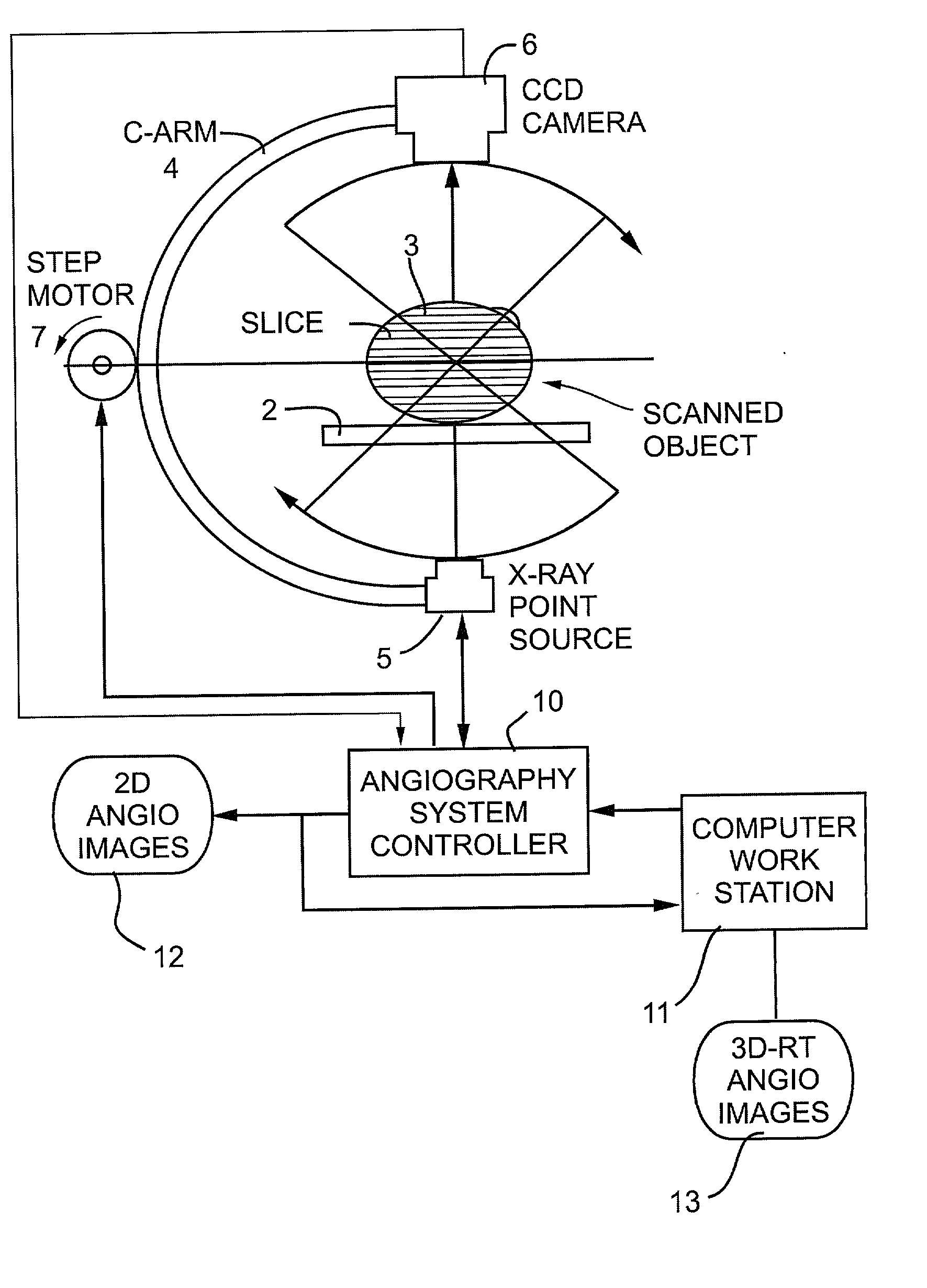
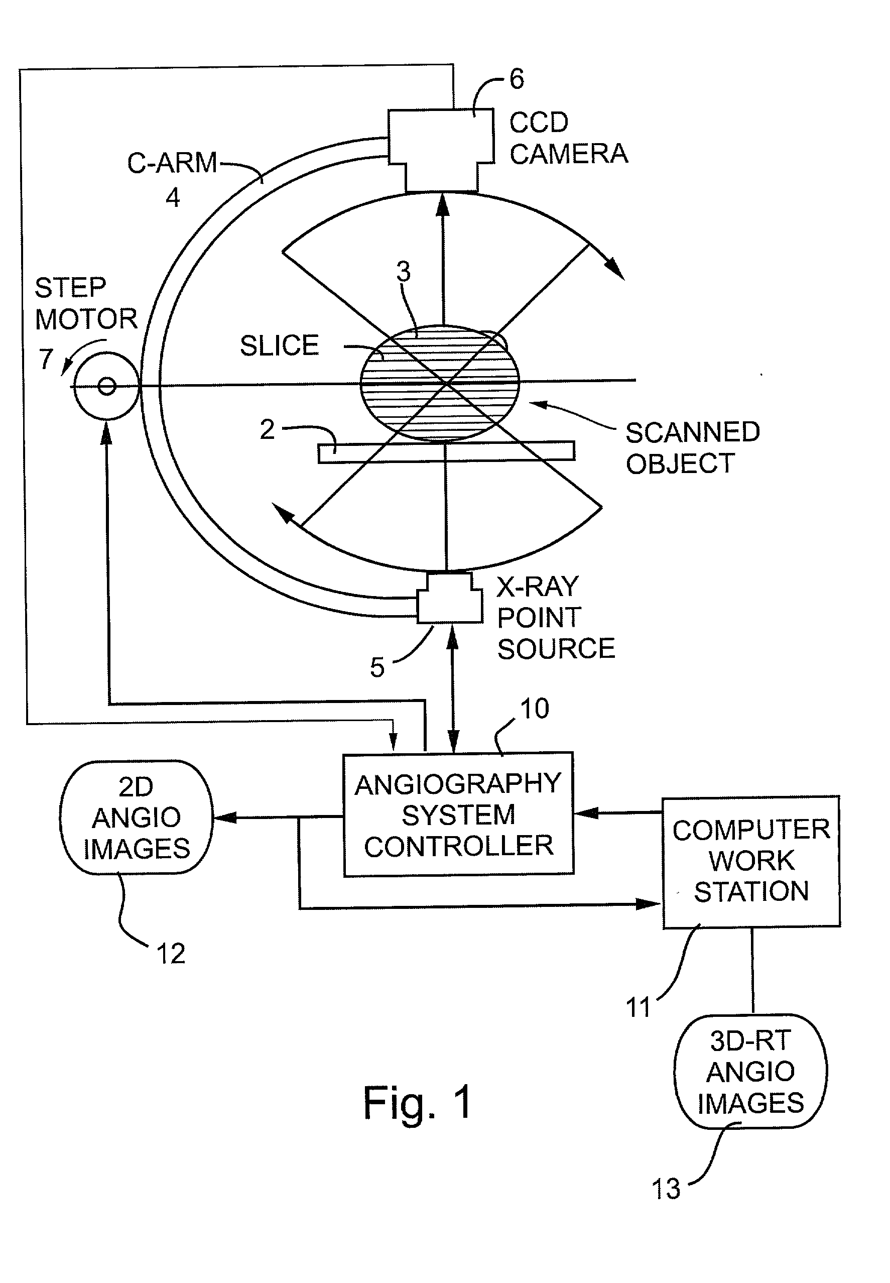
[0039] FIG. 1 schematically illustrates one form of apparatus constructed in accordance with the present invention particularly useful for producing either two-dimensional angiographs and / or three-dimensional angiographs of a patient's vascular system.

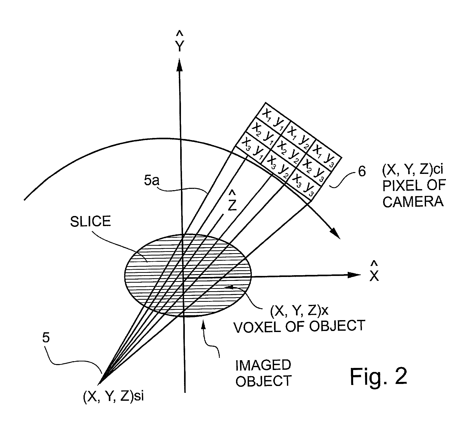
[0040] The system illustrated in FIG. 1 includes a horizontal support such as a table 2 for the patient 3 under examination, and a gantry C-arm 4, such as used in CT examination apparatus, enclosing the patient's body 3. The C-arm supports a radiation source 5 at one side of the patient's body, and a radiation detector 6 at the opposite side and in alignment with the radiation source. The radiation source 5 is an X-ray point source which produces a conical beam 5a as shown in FIG. 2; and the radiation detector 6, preferably a CCD camera, includes a two-dimensional matrix of detector elements as best seen in FIG. 4. As shown particularly in FIG. 2, the conical beam 5a produced by the radiation source 5 ...


PUM


Abstract

A method and apparatus for angiographically imaging a portion of a patient's vascular system, by: (a) producing a first sequence of masking images of the portion of the patient's vascular system; (b) determining the masking image in the sequence having a minimum change over its immediately preceding masking image in the sequence; (c) injecting a contrast material into the patient's vascular system; (d) producing a corresponding sequence of contrast images of the portion of the patient's vascular system while containing the contrast material and (e) subtracting from each contrast image of the sequence the corresponding masking image in the sequence starting with the minimum change masking image, to thereby produce a difference image having an enhanced contrast-to-noise ratio (CNR).

Description

[0001] The present application is a continuation-in-part of my prior U.S. application Ser. No. 09 / 946,168, filed Sep. 5, 2001, the total contents of which are incorporated herein by reference.FIELD AND BACKGROUND OF THE INVENTION[0002] The present invention relates to methods and apparatus for imaging two-dimensional (2-D) planes or surfaces and / or three-dimensional (3-D) objects. The invention is particularly useful in angiography for imaging a person's vascular system in 2-D or 3-D, and is therefore described below with respect to this application.[0003] Electromagnetic radiation has long been used to produce images of internal structures of the human body for purposes of diagnosis or treatment of diseases. One technique which has gained widespread use since the 1970's is computerized tomography (CT), sometimes called computerized axial tomography (CAT), in which a narrow beam of X-rays is swept across an area of the body and is recorded by a radiation detector as a pattern of ele...

Claims


Application Information

Patent Timeline
IPC IPC(8): A61B6/00
CPCA61B6/481A61B6/504
Inventor KEREN, HANAN
Owner MEDIMAG C V I
Why Eureka
Try Eureka