
Assaying ovarian cyst fluid

a technology of ovarian cysts and fluids, applied in the field of dna analysis, can solve the problems of serious complications in 15% of women, potentially lethal, and the inability to aspirate ovarian cyst fluid for cytology, and achieve the effect of not being a standard-of-care aspiration method

Active Publication Date: 2022-03-29
THE JOHN HOPKINS UNIV SCHOOL OF MEDICINE
View PDF332 Cites 1 Cited by
  • Summary
  • Abstract
  • Description
  • Claims
  • Application Information

AI Technical Summary

Benefits of technology

[0018]These and other embodiments which will be apparent to those of skill in the art upon reading the specification provide the art with powerful methods for assessing ovarian cysts without recourse to unnecessary surgeries.

Problems solved by technology

In addition to the anxiety that such findings provoke, many women undergo unnecessary surgery for cysts that are not malignant and may not be responsible for the symptoms they have.
Compounding this issue is the fact that surgery for ovarian cysts requires general anesthesia and is associated with significant morbidity, causing serious complications in 15% of women.
At the other end of the spectrum are epithelial ovarian cancers, which are potentially lethal and unequivocally require surgery.
However, aspiration of ovarian cyst fluid for cytology is not standard-of-care.
Because there is currently no reliable way to determine whether an ovarian cyst is malignant prior to surgical excision, many women undergo unnecessary, invasive surgeries for non-malignant lesions.

Method used


Image

  • Assaying ovarian cyst fluid
  • Assaying ovarian cyst fluid
  • Assaying ovarian cyst fluid

Examples


example 1

and Methods

Patient Samples

[0048]Cyst fluids were collected prospectively from 77 women presenting with a suspected ovarian tumor. Patients were diagnosed by transvaginal sonography or computed tomography and admitted for surgical removal of the cyst by gynecologic oncology surgeons at Sahlgrenska University Hospital, Gothenburg, Sweden. The study was approved by the ethical board of Gothenburg University and patients provided written consent. According to the approved protocol, ovarian cyst fluids were collected after removal of the cyst from the abdomen. All samples were immediately put in 4° C. for 15-30 minutes, centrifuged for 10 minutes at 500 g, and aliquoted into Eppendorf tubes. The fluids were transferred to −80° C., within 30-60 minutes after collection. All histology was reviewed by board-certified pathologists (Table 1).

[0049]Plasma HE4 concentrations were determined using a commercial HE4 EIA assay (Fujirebio Diagnostics) and plasma CA-125 levels were measured using the...

example 2

istics of the Tumors and Cyst Fluid Samples

[0053]DNA was isolated from surgically excised ovarian cysts of 77 women. Ten of them had non-neoplastic cysts, 12 had benign tumors, 24 had borderline tumors, and 31 had cancers (13 Type I and 18 Type II). Age, histopathologic diagnosis, stage, and other clinical information are provided in Table 1. The median amount of DNA recovered from the cysts was 222 ng (interquartile range (IQR) of 53 to 3120 ng) (Table 2). There was no significant difference in the amounts of DNA between borderline tumors and type I or type II cancers (Table 2). However, the borderline tumors and cancers contained significantly more DNA than the non-neoplastic cysts or benign tumors (4453±6428 ng vs. 62±64 ng; p<0.001, Wilcoxon rank-sum test).

example 3

ex PCR-Based Test to Identify Tumor-Specific Mutations in Cyst Fluid Samples

[0054]We designed a multiplex PCR-based test that could simultaneously assess the regions of 17 genes frequently mutated in ovarian tumors. The amount of DNA shed from neoplastic cells was expected to be a minor fraction of the total DNA in the cyst fluid, with most DNA emanating from normal cells. We therefore used a sensitive detection method, called Safe-SeqS (Safe-Sequencing System), to identify mutations in cyst fluid samples (34). In brief, primers were designed to amplify 133 regions, covering 9054 distinct nucleotide positions within the 17 genes of interest (Table S1). Three multiplex PCR reactions, each containing non-overlapping amplicons, were then performed on each sample. One primer in each pair included a unique identifier (UID) for each template molecule, thereby drastically minimizing the error rates associated with PCR and sequencing, as described previously (34) (Table S1). Under the condi...


PUM

PropertyMeasurementUnit
areaaaaaaaaaaa
mutant allele frequencyaaaaaaaaaa
physicalaaaaaaaaaa

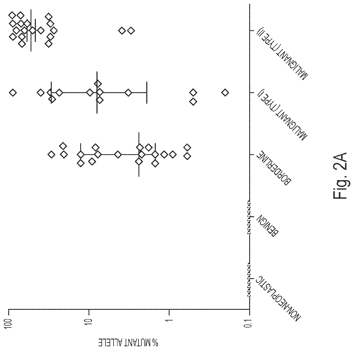
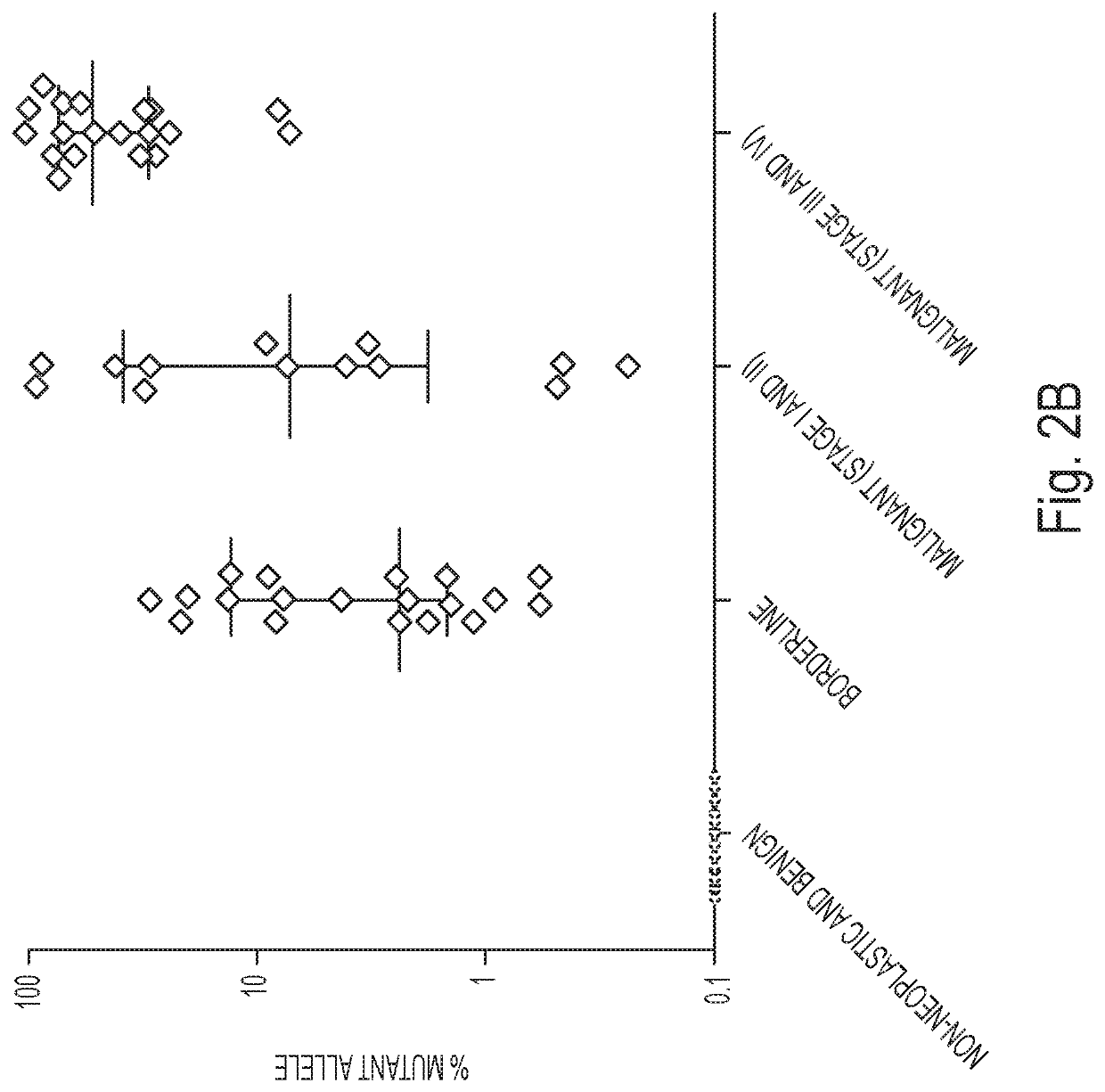
Abstract

A diagnostic test for ovarian cysts is based on the detection of mutations characteristic of the most common neoplasms giving rise to these lesions. With this test, tumor-specific mutations were detected in the cyst fluids of 19 of 24 (79%) borderline tumors and 28 of 31 (90%) malignant ovarian cancers. In contrast, we detected no mutations in the cyst fluids from 10 non-neoplastic cysts and 12 benign tumors. When categorized by the need for exploratory surgery (i.e., presence of a borderline tumor or malignant cancer), the sensitivity of this test was 85% and the specificity was 100%. These tests could inform the diagnosis of ovarian cysts and improve the clinical management of the large number of women with these lesions.

Description

[0001]This application is a National Stage application under 35 U.S.C. § 371 of International Application No. PCT / US2016 / 046453, having an International Filing Date of Aug. 11, 2016, which claims the benefit of U.S. Provisional Application Nos. 62 / 203,573, filed Aug. 11, 2015, each of which is incorporated herein by reference in its entirety.[0002]This invention was made with government support under CA 43460, 57345, and 62924 awarded by National Institutes of Health. The government has certain rights in the invention.TECHNICAL FIELD OF THE INVENTION[0003]This invention is related to the area of DNA analysis. In particular, it relates to analysis of genes in clinical samples.BACKGROUND OF THE INVENTION[0004]Ovarian cancer is the most lethal gynecologic malignancy, with 21,980 estimated new cases and 14,270 estimated deaths in the United States in 2014. Approximately 1.3% of women will be diagnosed with ovarian cancer during their lifetime (1). These cancers commonly present as an ad...

Claims


Application Information

Patent Timeline
Patent Type & Authority Patents(United States)
IPC IPC(8): C12Q1/6886
CPCC12Q1/6886C12Q2600/156
Inventor WANG, YUXUANVOGELSTEIN, BERTKINZLER, KENNETH W.DIAZ, LUISPAPADOPOULOS, NICKOLASSUNDFELDT, KARINKRISTJANSDOTTIR, BJORG
Owner THE JOHN HOPKINS UNIV SCHOOL OF MEDICINE
Learn More